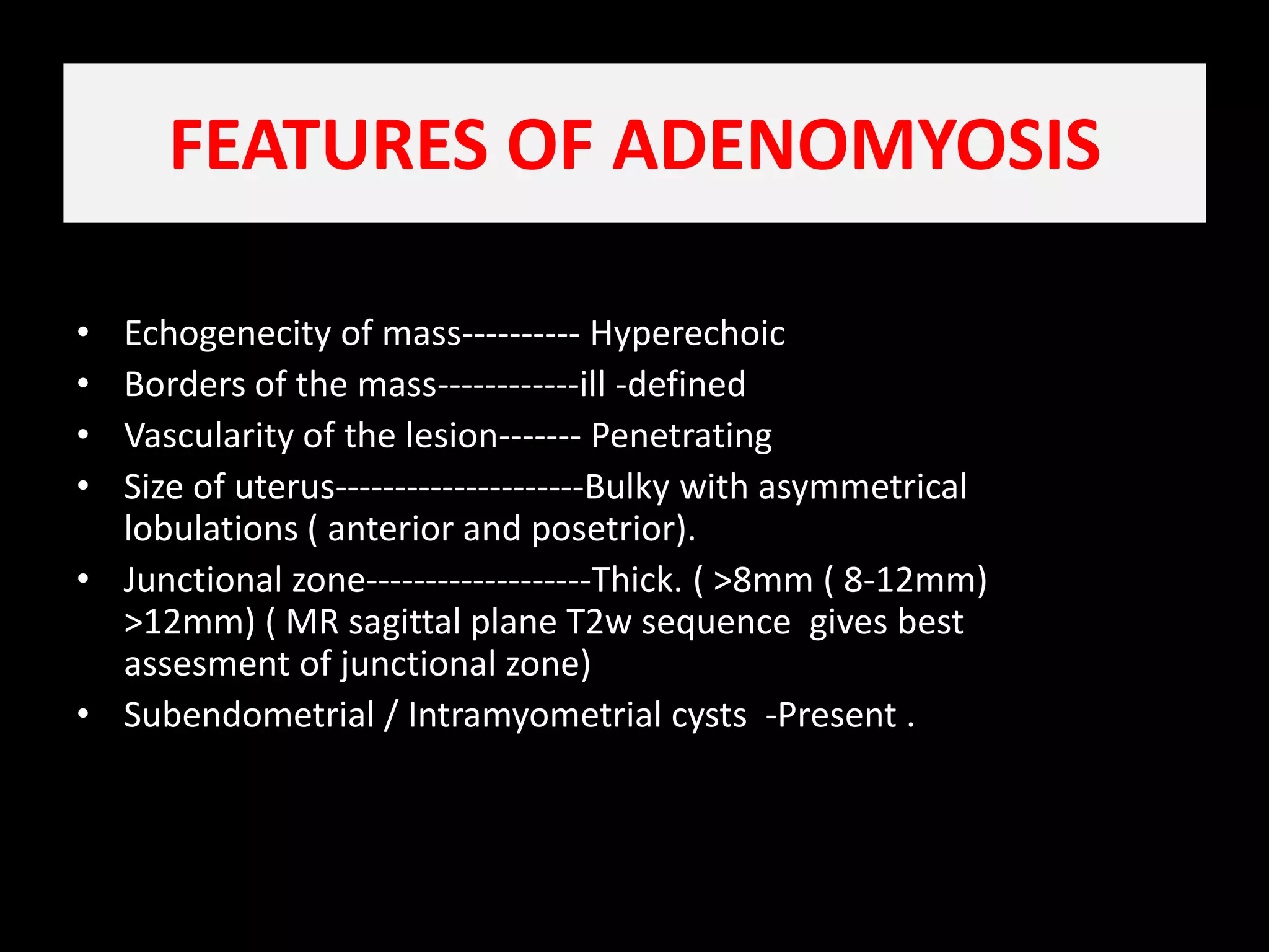
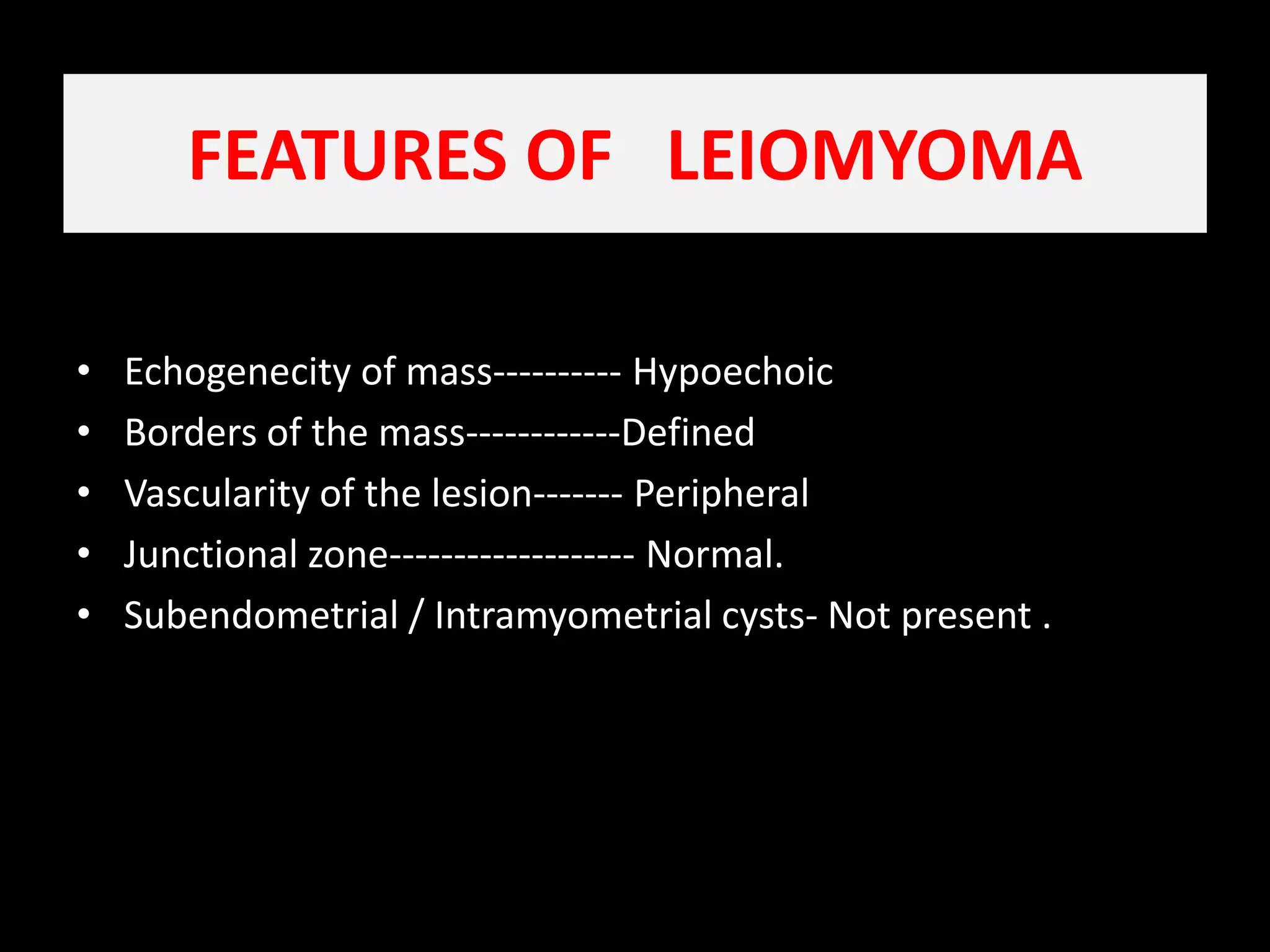
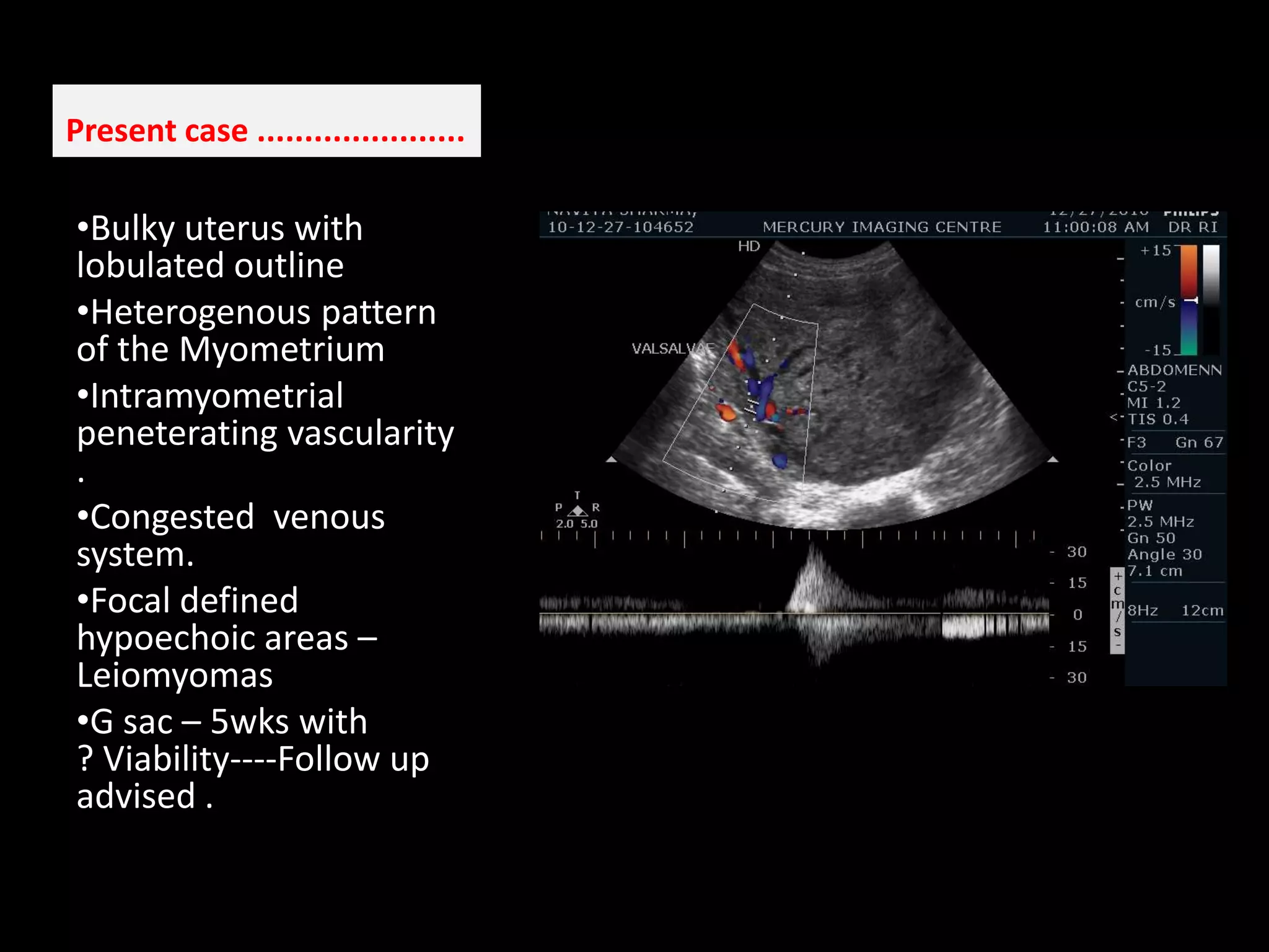
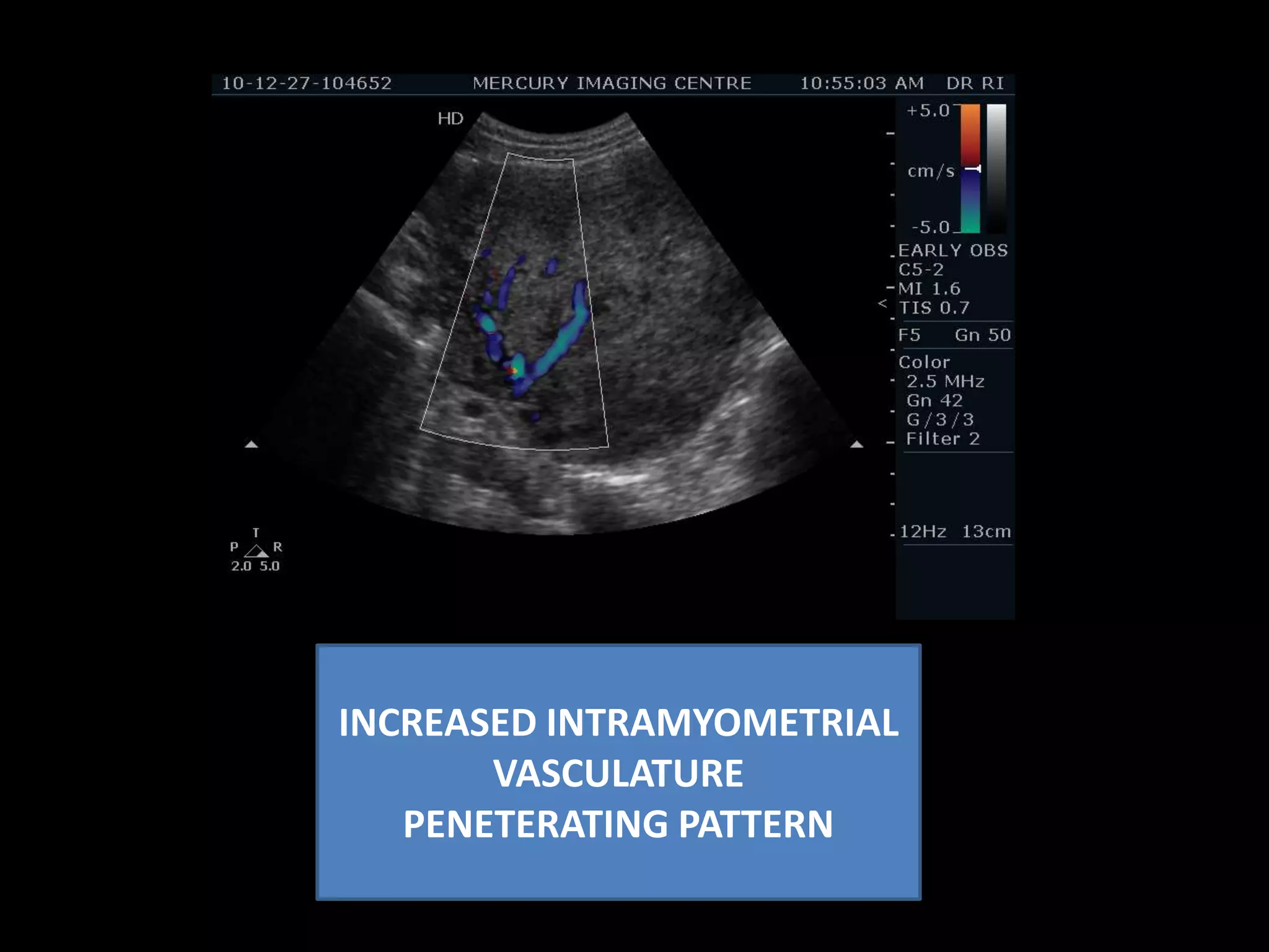
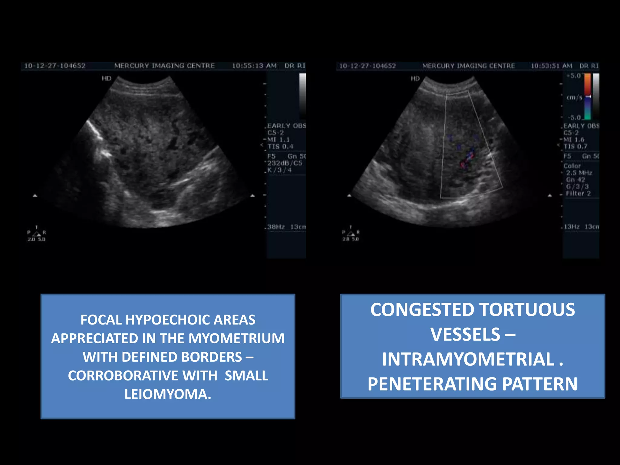
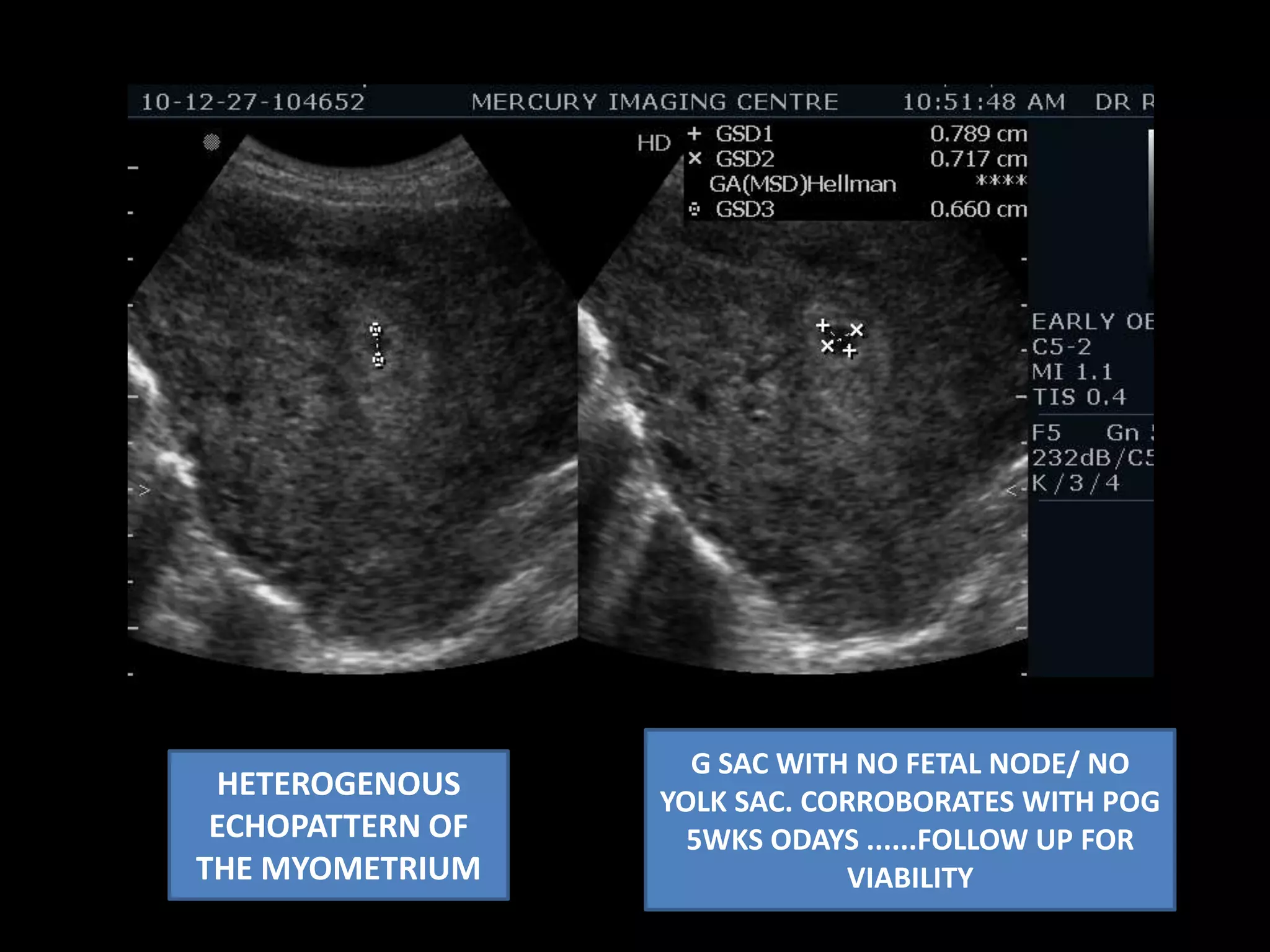
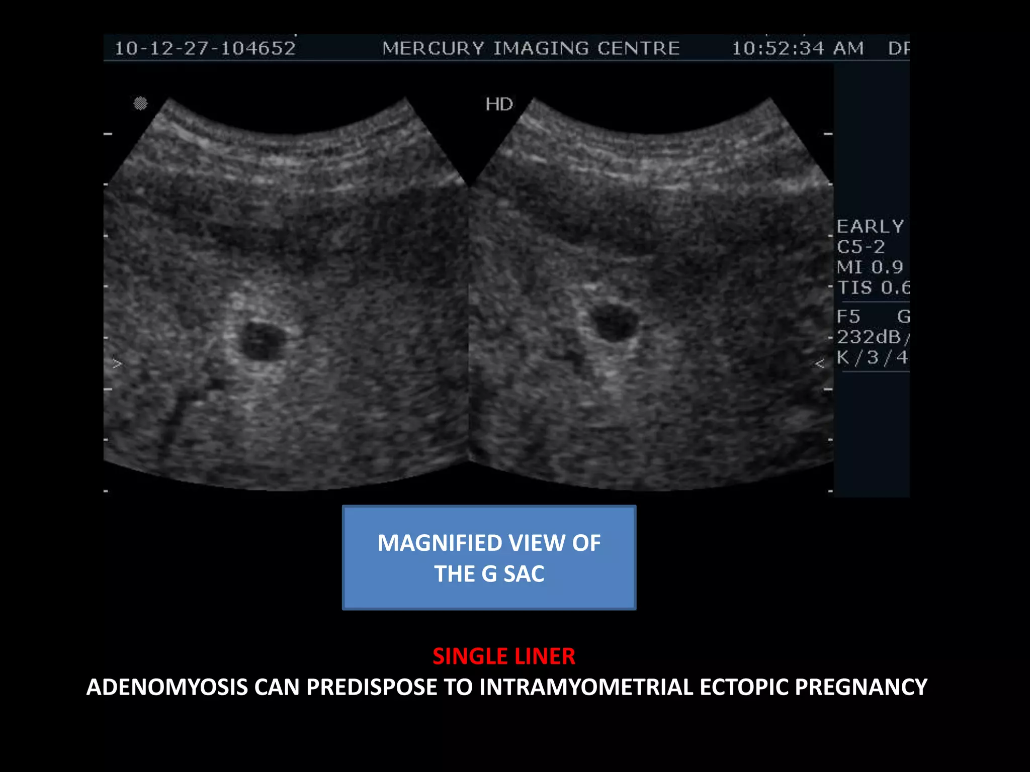
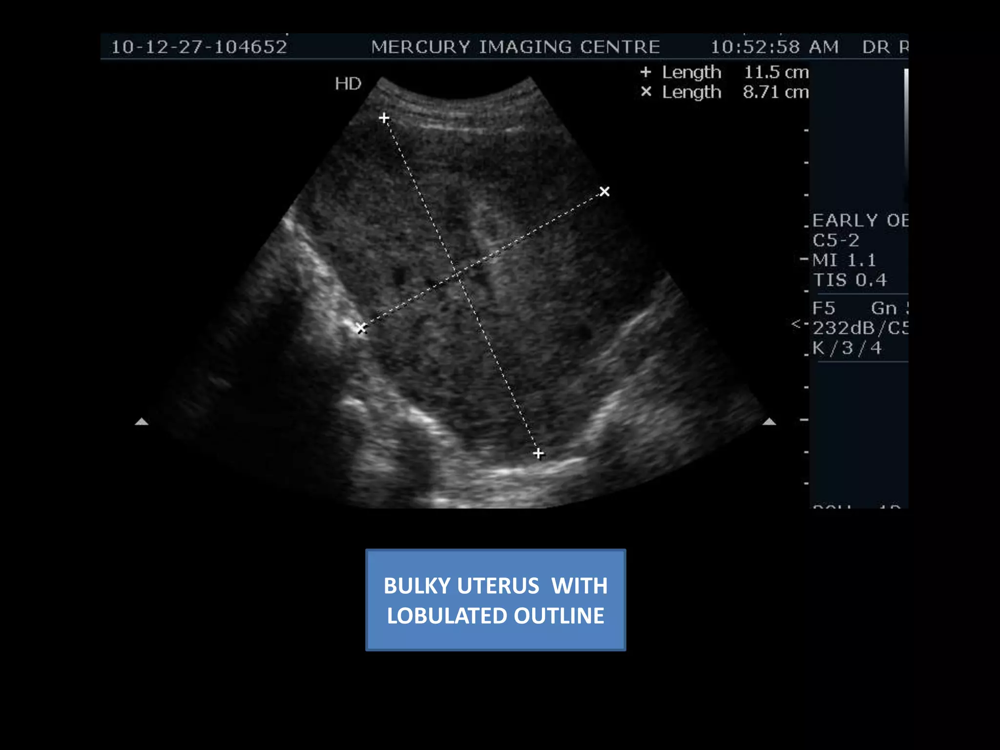
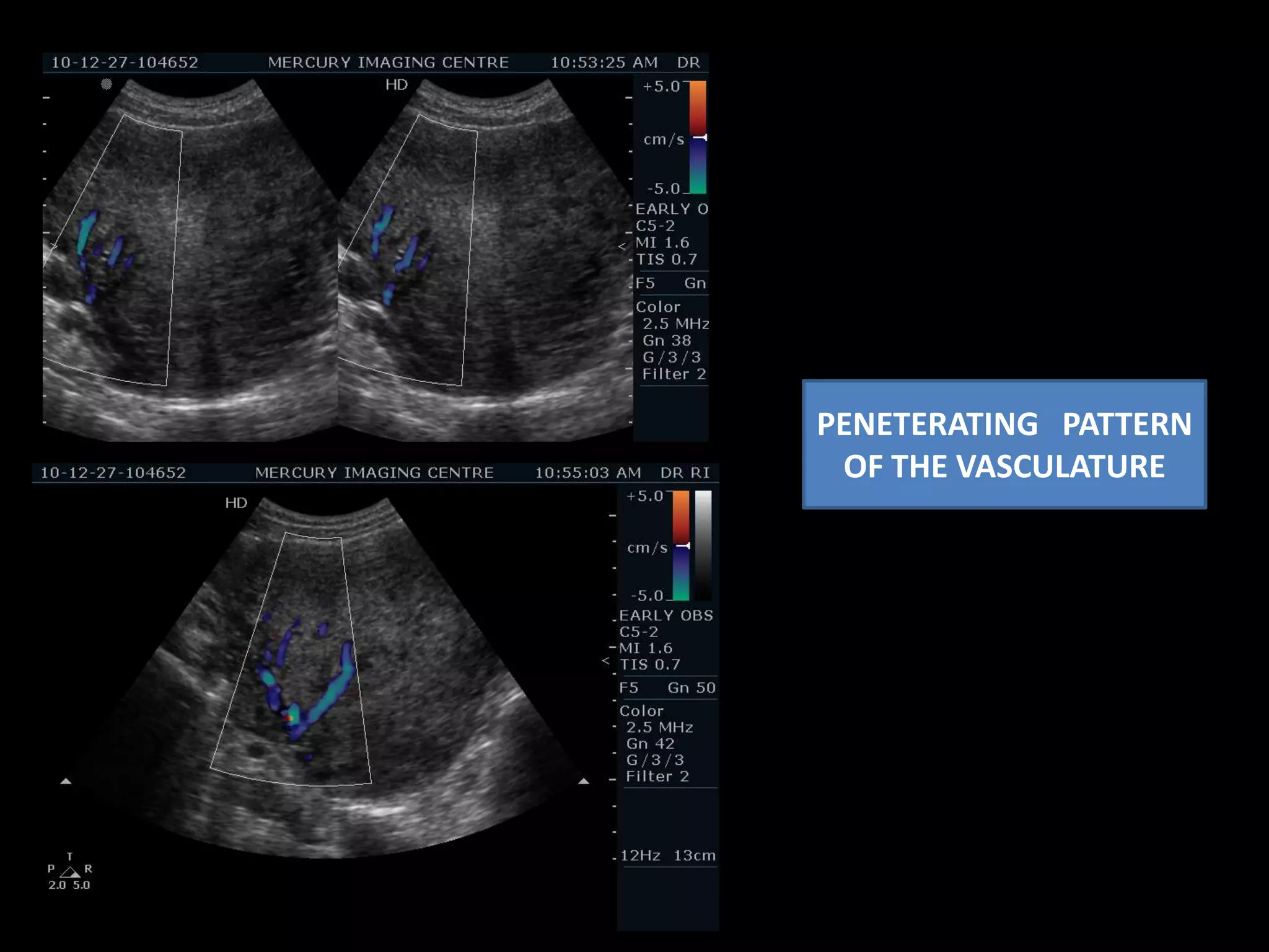
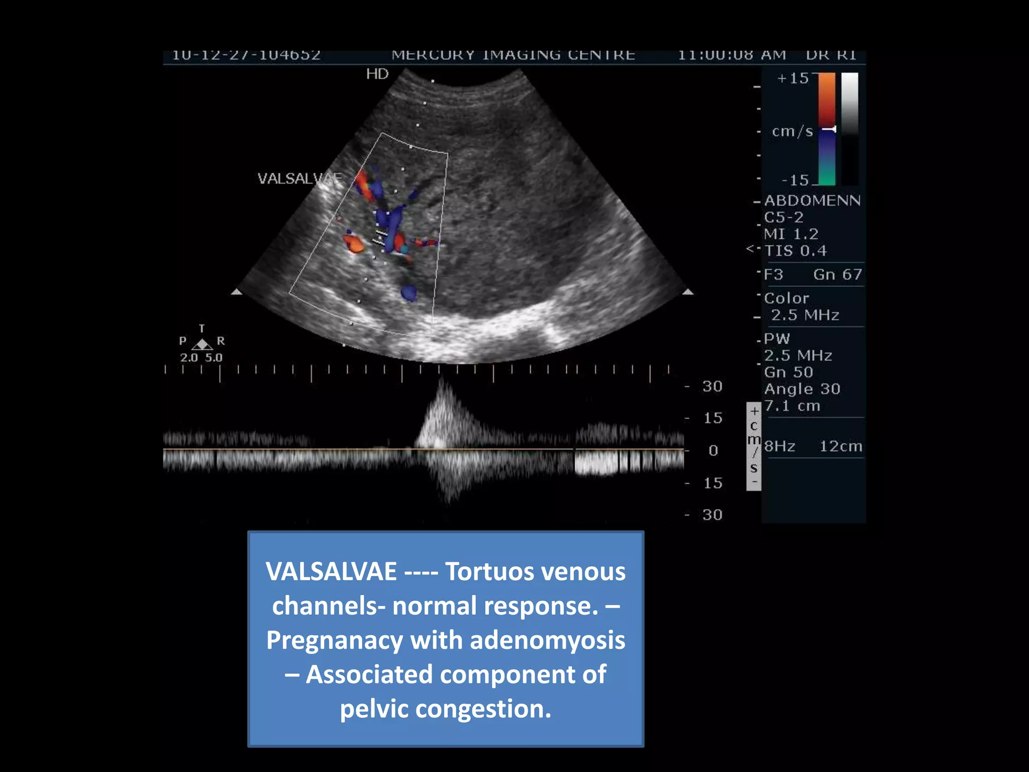
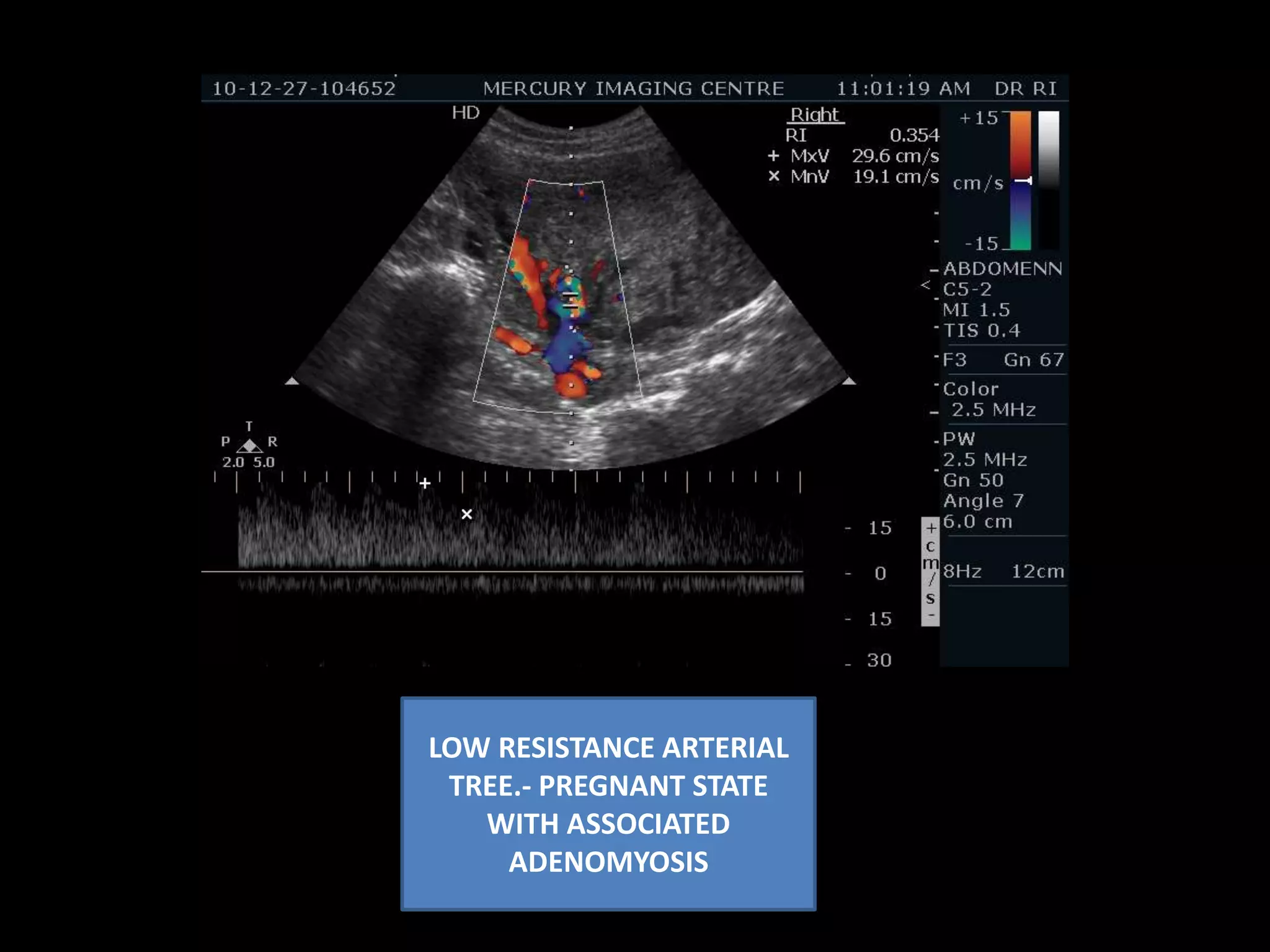
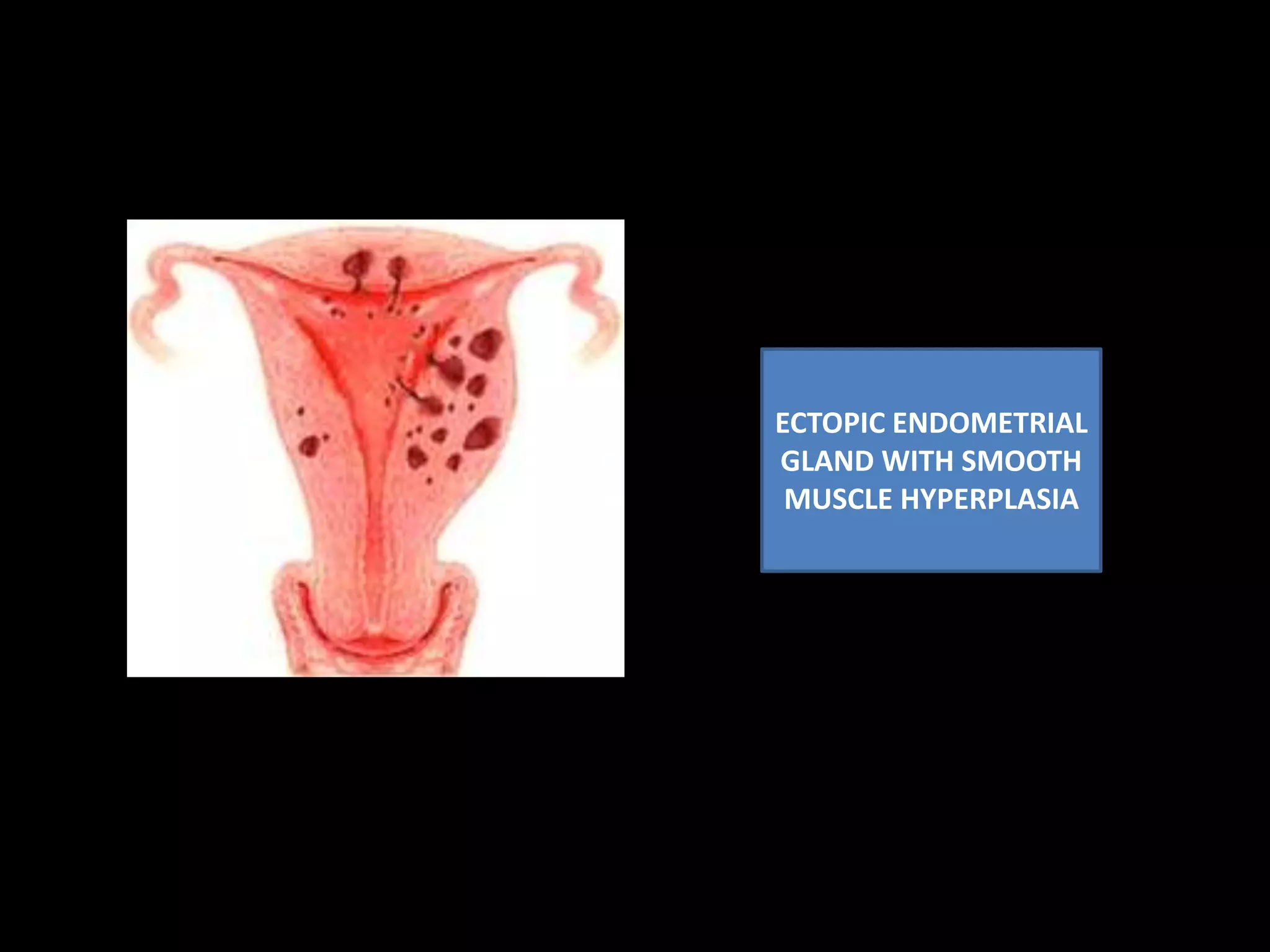
A 22-year-old female presented with episodic lower abdominal pain and a pregnancy of 5 weeks. Ultrasound findings included a bulky uterus with lobulated outlines and heterogeneous myometrium. Increased penetrating intramural vascularity and congested tortuous vessels were seen. Focal hypoechoic areas with defined borders represented small leiomyomas. A gestational sac was seen but follow up was needed to assess viability. These findings were consistent with adenomyosis. Adenomyosis can be differentiated from leiomyoma based on features such as echogenicity, borders, vascularity, and presence of subendometrial cysts.


![Rheumatic Fever CASE PRESENTATION [Autosaved].pptx](https://cdn.slidesharecdn.com/ss_thumbnails/casepresentationautosaved-251123182512-9d9b0da4-thumbnail.jpg?width=640&height=640&fit=bounds)


