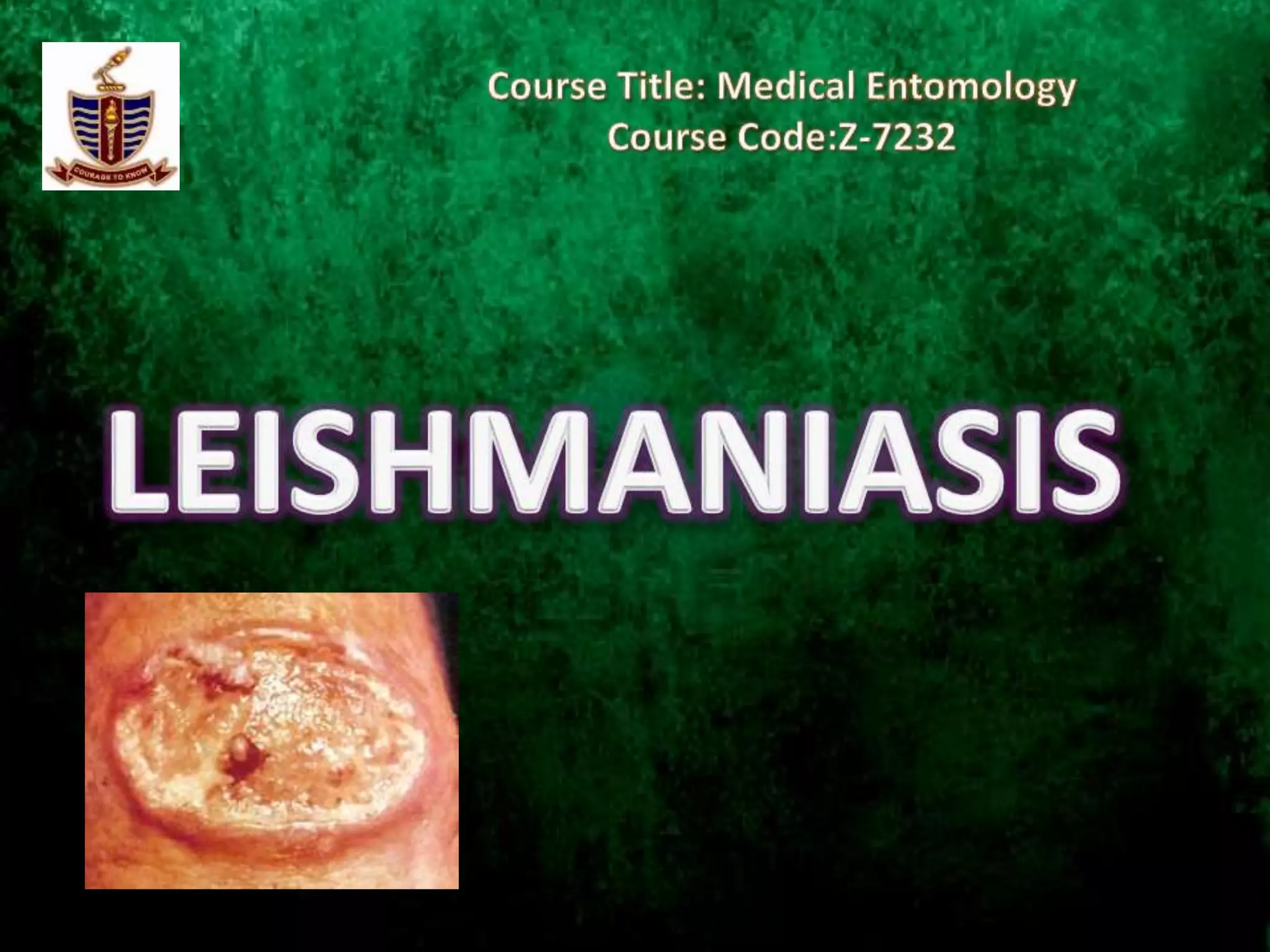
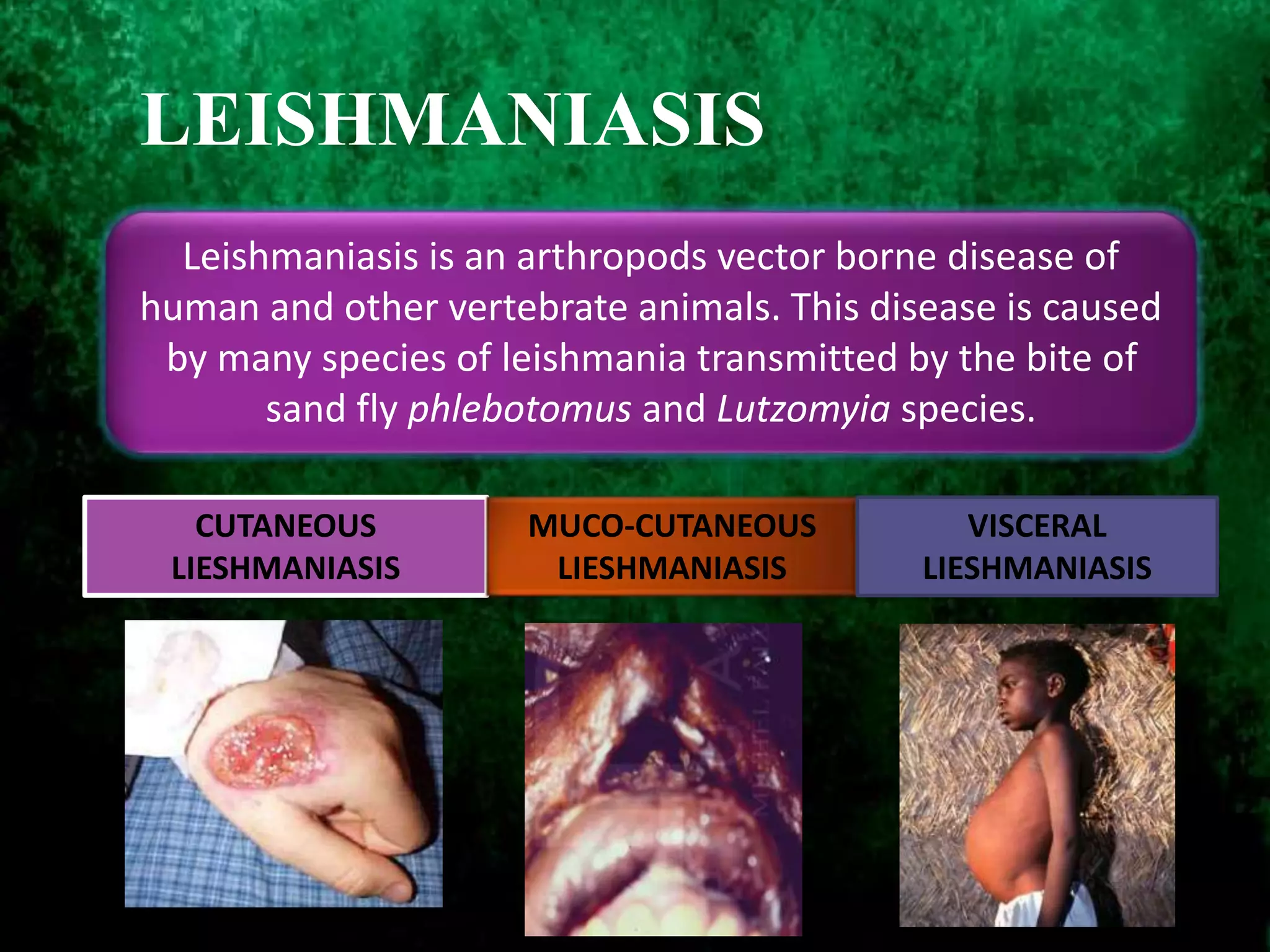
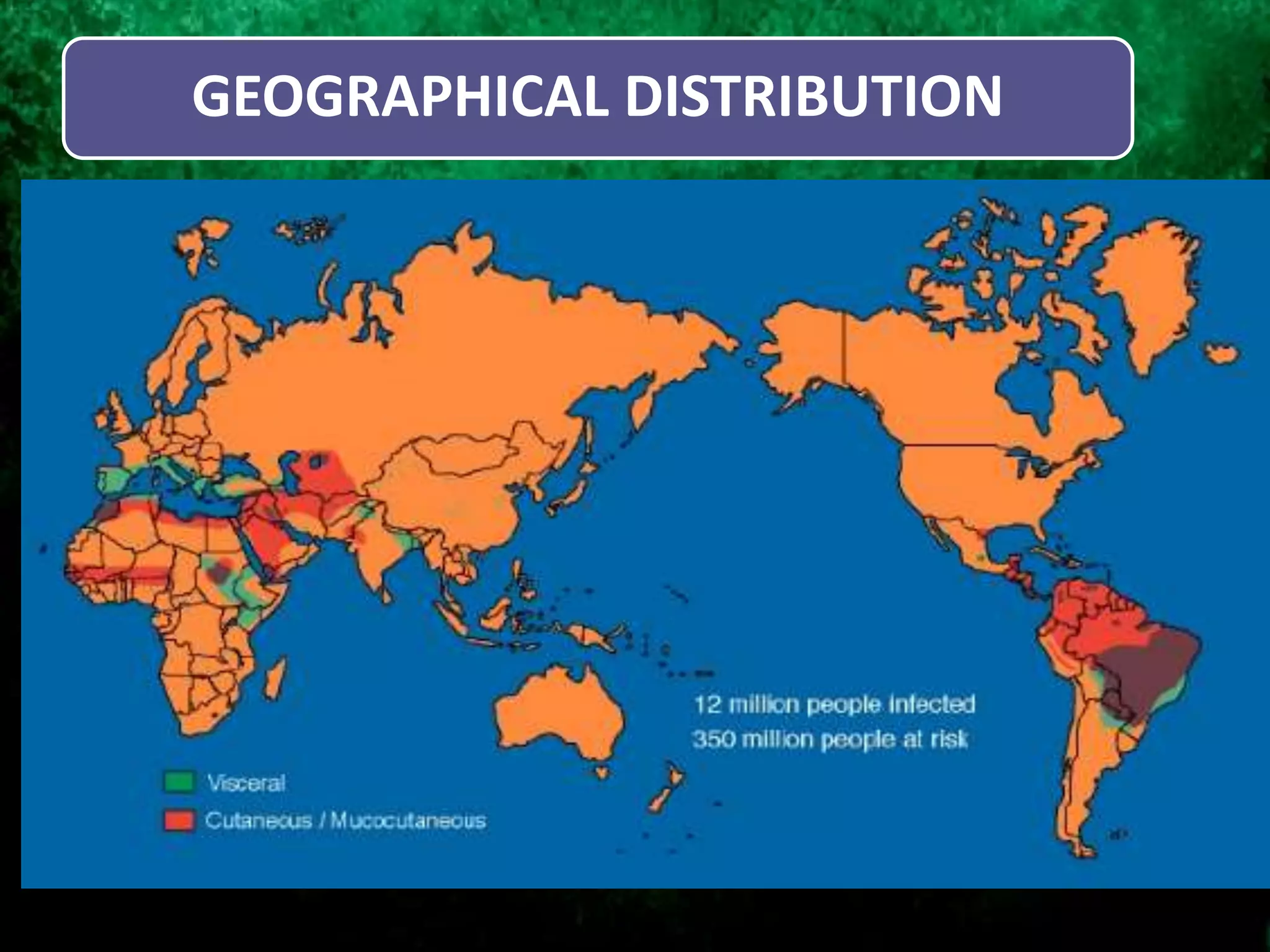
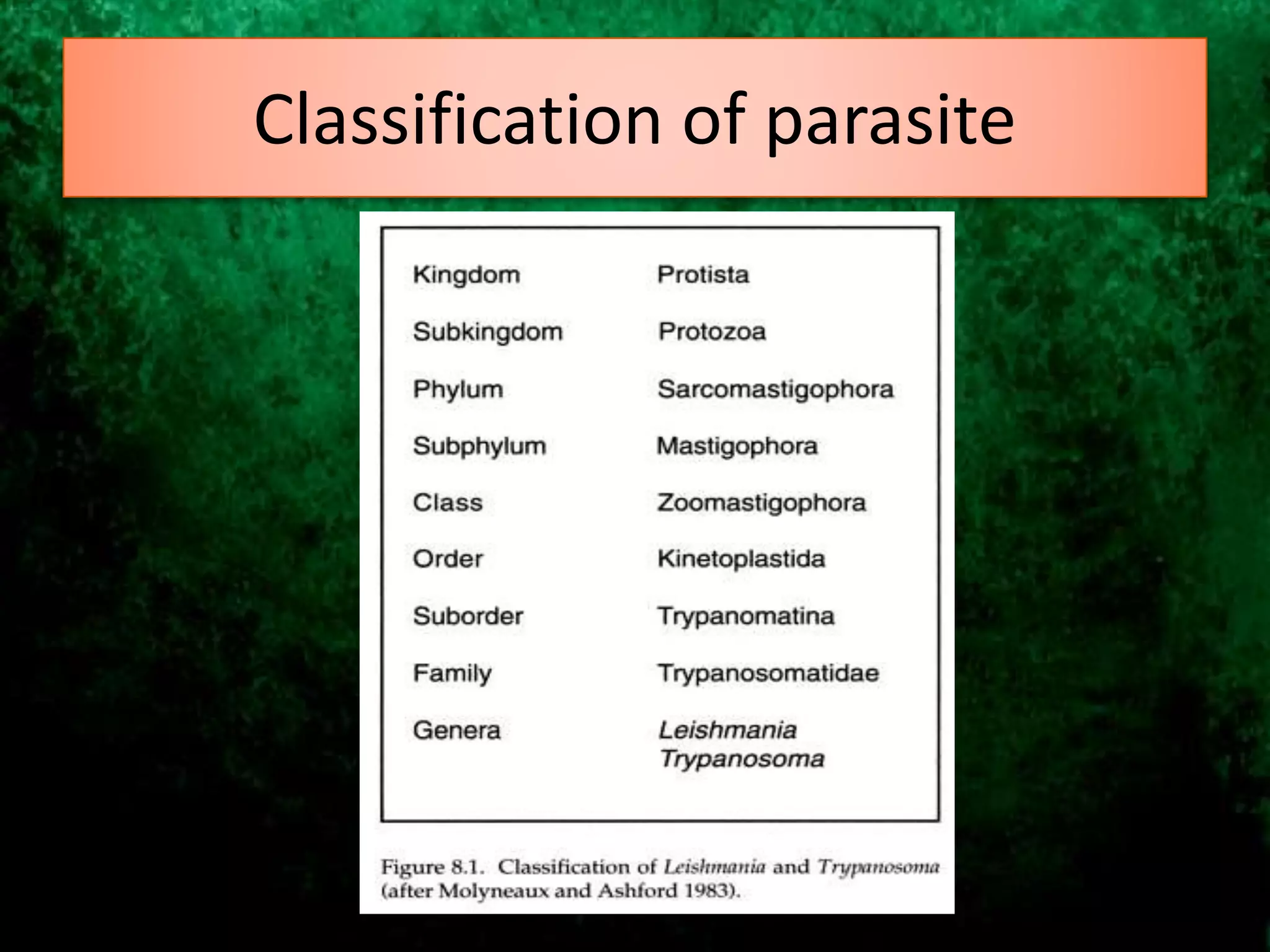
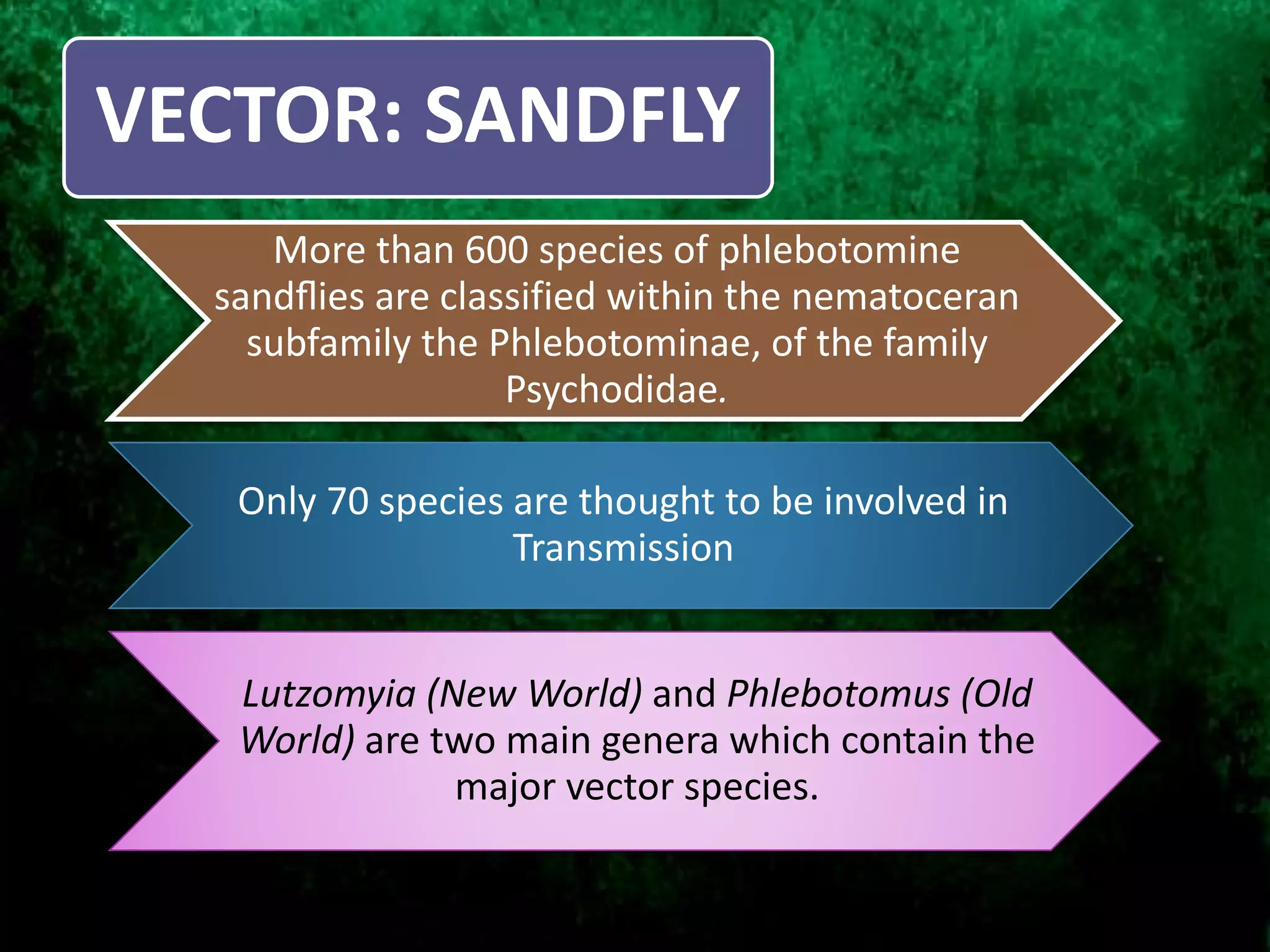
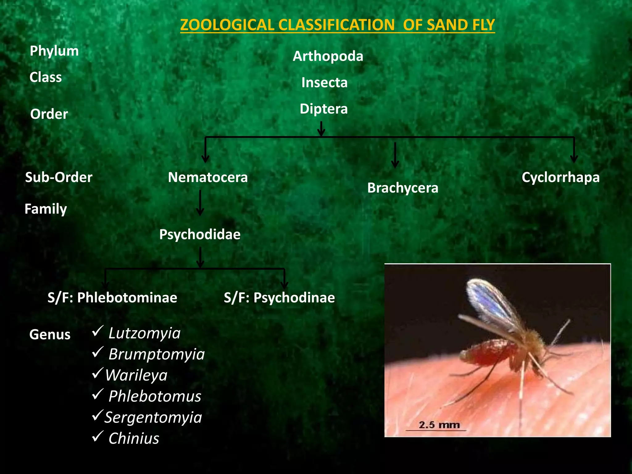
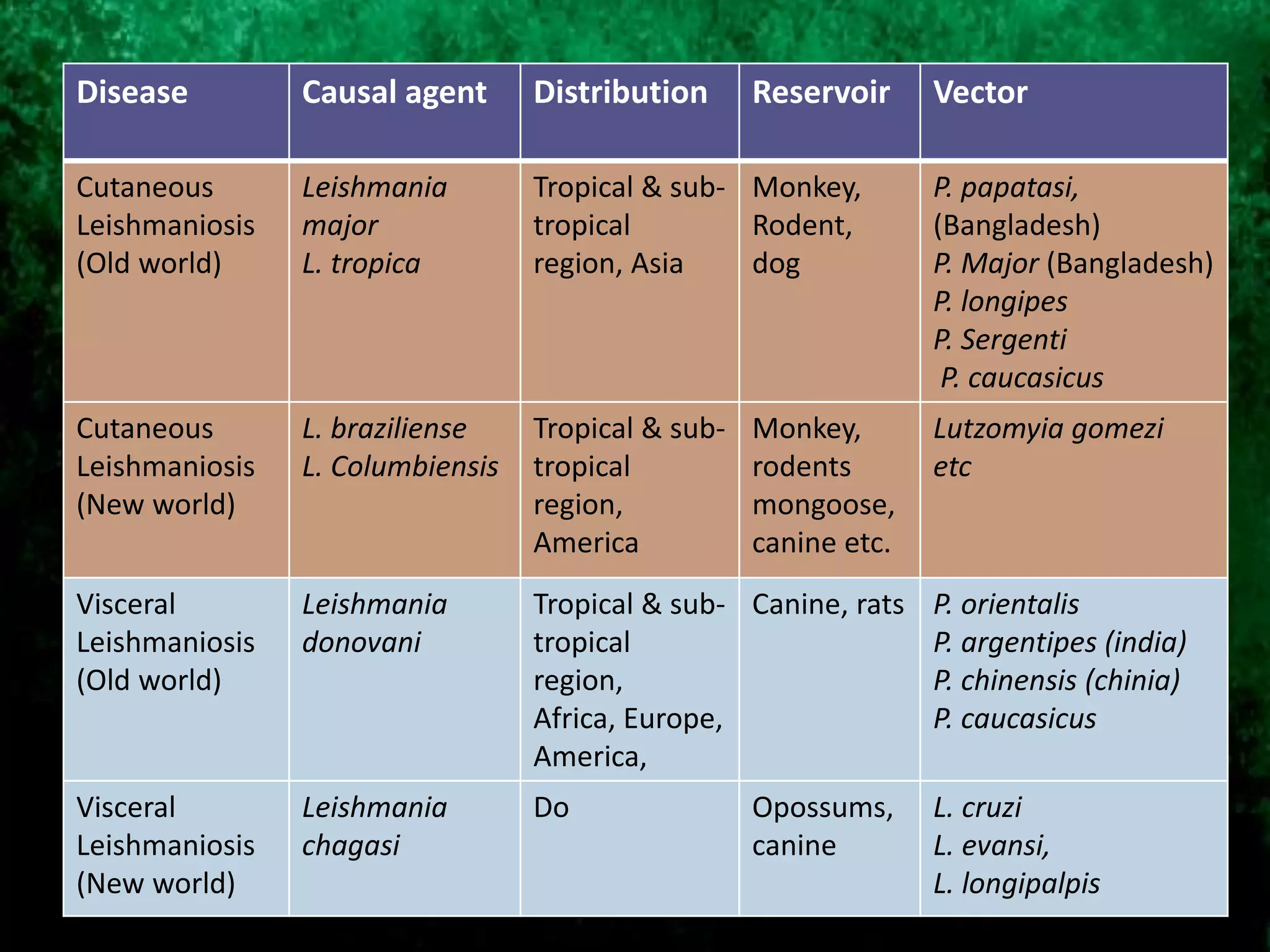
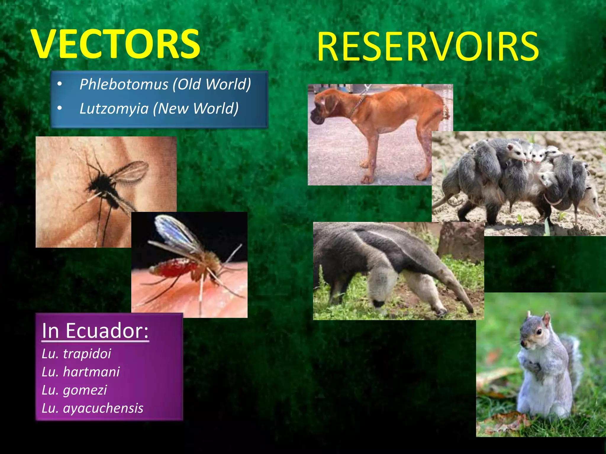
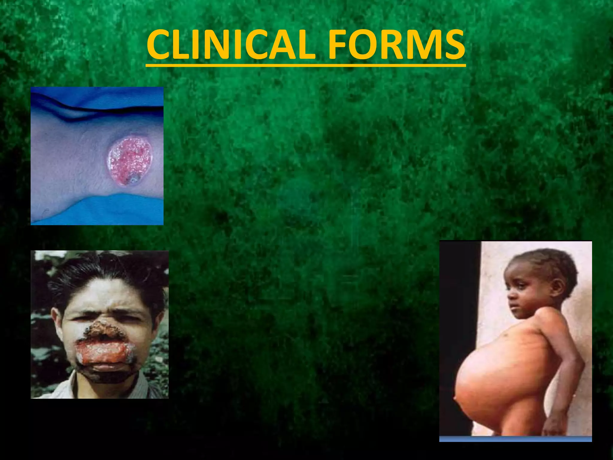
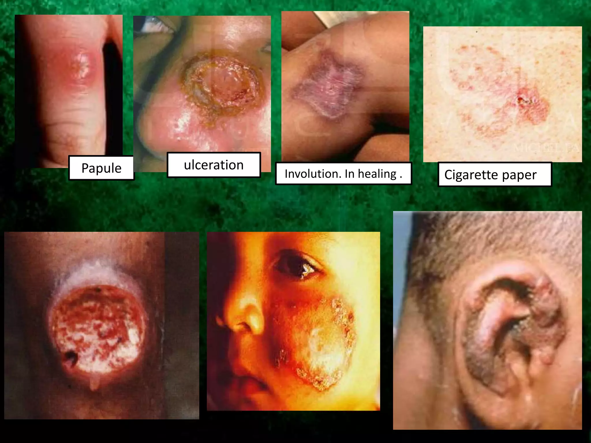
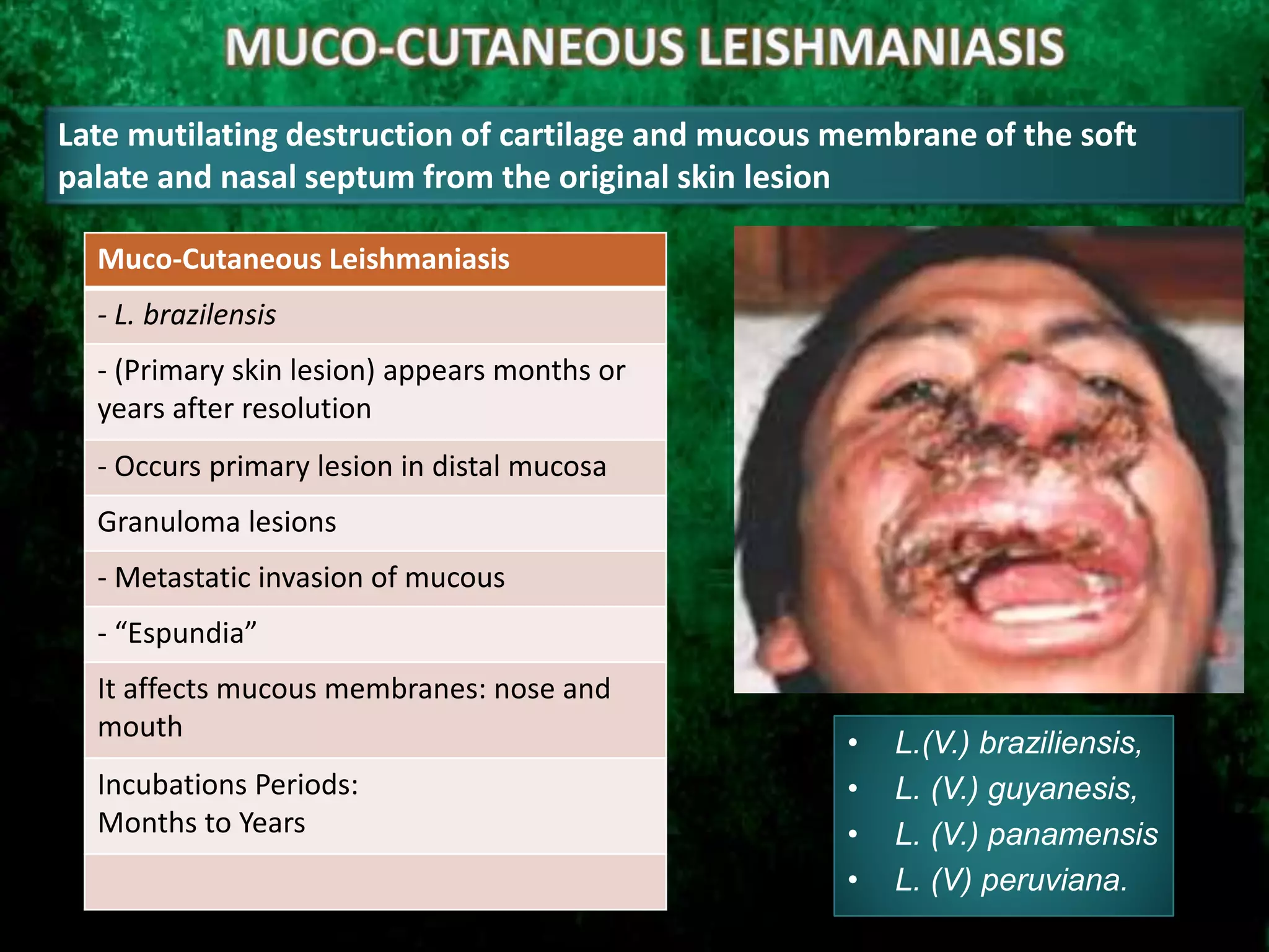
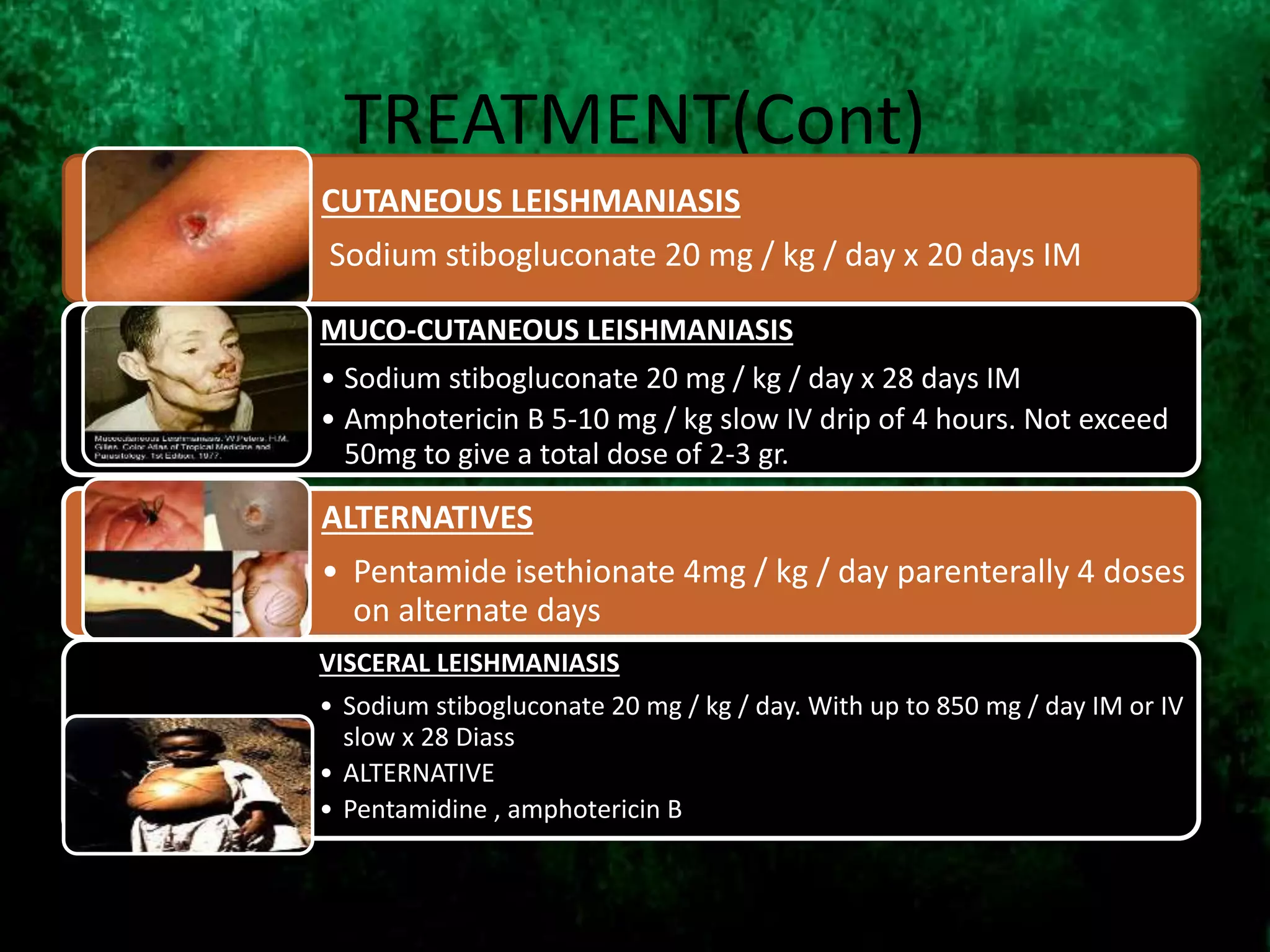
Governament College University Lahore
Zoology Department
SARWAR ALLAH DITTA
ROLL # 657
Leishmaniasis is an arthropods vector borne disease of
human and other vertebrate animals. This disease is caused
by many species of leishmania transmitted by the bite of
sand fly phlebotomus and Lutzomyia species.
CUTANEOUS
LIESHMANIASIS
MUCO-CUTANEOUS
LIESHMANIASIS
VISCERAL
LIESHMANIASIS
LEISHMANIASIS
Found in parts of at least 88 countries including the
Middle East
In the Middle East L. major and L. tropica are the
most common species
• L. major causes skin infection
• L. tropica causes skin and visceral infection and rarely causes
mucocutaneous infection
12 million people
infected worldwide.
Tropical and subtropical
regions.
In Latin America the
disease is rural .
Vectors and reservoirs
are located in
geographic areas.
In Ecuador endemic in
mountainous and
jungle areas of coast
and mountains .
Disease outbreaks
related to climatic or
demographic changes.
In Africa and Southern
Europe 70 % of
individuals with
leishmaniasis have AIDS
.
500,000 new cases of
visceral leishmaniasis
estimated to occur each
year.
In Africa and Southern
Europe 70 % of
individuals with
leishmaniasis have AIDS
.
EPIDEMIOLOGY
GEOGRAPHICAL DISTRIBUTION
Geographical Distribution(Cont)
Leishmania species have been reported almost on every continent
of the world.
Human populations are mainly affected by this disease
Africa, parts of Asia, the Middle East, Latin America and the
Mediterranean regions
Severity and type of disease is mainly subjected to the endemic
species of leishmania in that particular region (Banuls, et al., 2007).
Leishmaniasis in the Middle East
90% of cutaneous leishmaniasis occurs in
Afghanistan, Iran, Saudi Arabia, Syria, Brazil and Peru
• 8,779 cases were reported in Iraq in 1992
• Sore is commonly called the Baghdad boil
90% of all visceral leishmaniasis occurs in
Bangladesh, Brazil, India, and the Sudan
• 2893 cases were reported in Iraq in 2001
90% of mucocutaneous leishmaniasis occurs in
Bolivia, Brazil and Peru
• Rarely associated with L. tropica which is found in Middle East
Geographical Distribution(Cont)
In South Asia and Africa L. donovani causes VL.
L. infantum causes VL in the Mediterranean, the Middle East, Latin America and parts of Asia (Herwaldt,
1999).
L. major causes CL in Africa, the Middle East and parts of Asia.
L. tropica causes CL in Middle East, the Mediterranean and parts of Asia.
L. aethiopica causes CL in some parts of Africa.
Different species cause CL from Mexico through South America.
L. Mexicana complex cause CL in south Central Texas.
Australia was free of leishmania species until 2004. Now some species are reported in Kangroos, Wallabies,
and other marsuipials.
Classification of parasite
Classification
Viannia Leishmania
Complex L. (V ) braziliensis Species:
L. (V.) braziliensis
L. (V.) peruviana
Complex L. (V ) guyanensis Species:
L. (V.) guyanensis
L. (V.) panamensis
L. (V.) shawi
Complex L. (V ) lainsoni Species:
L. (V.) lainsoni
Complex L. (V ) Naiff Species:
L. (V.) naiffi
Complex Leishmania (Leishmania) mexicana
• L. (L.) mexicana
Complex Leishmania (Leishmania) amazonensis
• L. (L.) amazonensis
Complex Leishmania (Leishmania) infantum
• L. (L.) infantum
Complex Leishmania (Leishmania) donovani
• L. (L.) donovani
• L. (L.) Archibaldi
Complex Leishmania (Leishmania) trópica
• L. (L.) trópica
Complex Leishmania (Leishmania) major
• L. (L.) major
• L. (L.) major-like
Complex Leishmania (Leishmania) aethiopica
• L. (L.) aethiopica
Sub Genera
Classifications of Leishmania Spp.
Leishmania L. major complex (L. major)
Old World
L. tropica complex (L. tropica, L. killicki)
L. aethiopica complex (L. aethiopica)
L. donovani complex (L. donovani, L. infantum)
Viannia L. donovani complex (L. chagasi)
New WorldL. mexicana complex
(L. mexicana, L. venezuelensis, L. garnhami,
L. amazonensis, L.pifanoi)
L. braziliensis complex (L. braziliensis, L. peruviana, L.
columbiensis, L. lainsoni)
L. guyanensis complex (L. guyanensis, L. panamensis)
VECTOR: SANDFLY
More than 600 species of phlebotomine
sandflies are classified within the nematoceran
subfamily the Phlebotominae, of the family
Psychodidae.
Only 70 species are thought to be involved in
Transmission
Lutzomyia (New World) and Phlebotomus (Old
World) are two main genera which contain the
major vector species.
Diptera
Nematocera
Brachycera
Cyclorrhapa
Arthopoda
Insecta
Order
Sub-Order
Family
Class
Phylum
Psychodidae
S/F: Phlebotominae S/F: Psychodinae
 Lutzomyia
 Brumptomyia
Warileya
 Phlebotomus
Sergentomyia
 Chinius
Genus
ZOOLOGICAL CLASSIFICATION OF SAND FLY
Family: Psychodidae
Psychodids:
 small nematoceran flies
 Long antennae
 Pendulous palps
 hairy bodies and wings
 wing venation: more or less parallel
Longitudinal veins, radial vein 3 or 4 branched and the
media 4 branched
Antennae: 16 segment
Pliose type (in male, female)
Palp: recurved, hairy , 5 segmented, 3rd segment bears sensilia
Old world species: 5th palpal segment longest
New old species: 3rd palpal segment longest
Phlebotomus spp (sand Fly)
Psychoda spp (Moth
Fly)
VECTOR: SANDFLY(Cont)
Adult sandflies are small
(1.5–4.0mm long),fragile,
slender-bodied, hairy,
brownish insects with long,
stilt-like legs. (Lehane, 2005)
At rest a pair of narrow,
lanceolate wings almost in
erect positions over the body
in the “V” like appearance.
Female Mouthparts are adapted for blood feeding and contains six blade like
stylet within a labial sheats.
VECTOR: SANDFLY (Cont)
Some species can produce one
clutch of eggs before their first
blood meal (Autogenous /Partly
Autogenous) e.g. Phlebotomus
papatasi
Some species need a blood meal
before they can produce any
eggs batch (Anautogenous)
(Lehane, 2005)
Most of them are exophagic, while a few
of them are endophagic (Lehane, 2005)
One blood meal can support the
production of about 100 eggs
Life Cycle of Sandfly
The life cycle of Sandfly is characterized by complete
metamorphosis, having 4 developmental stages.
Egg Stage
Larvae Stage (Four Instar)
Pupae Stage
Adult Stage
Life Cycle of Sandfly
Eggs Stage: The eggs are laid less than 100 in
number, and are brown or black in color. The
eggs size measure about 0.4 mm in length.
Eggs hatch almost in 1-2 weeks in moist
conditions
Larval Stage: is maggot like structure, having
large head, thorax and abdomen and two long
bristle on last abdominal segment. 12-
segmented larvae mainly feed on organic debris.
Four instars of larvae follow over a period of 21-
60 days (Peters, 1992) / 16-90 days (Service,
2001a).
Adult Stage: The average life span of
an adult sandfly is about 2 weeks
Pupae Stage: Pupae keep the skin of the
final larval stage with its characteristic
double pair of caudal bristles and can be
easily recognized. The pupal stage is
completed in less than two weeks. (Lehane.,
2005)
Bionomics of Phlemotominae
• Distribution:
 Old world phlebotomines are savanna and desert species ( low rainfall)
 Population increases after rainy seasons
 but P. papatasi and P. argentipes (India): seen in monsoon and seen in dry season
 New world phlebotomines are mainly inhabitants of forests ( high rain fall)
Breeding place places: dark, cool, humid niches like cracks, crevices of
soil, manure, rocks, tree hollows, nests, tree crotches, animal burrows,
(e.g. burrow of rodent), stables, livestock pens, well or any moist places
Host Range: Mostly broad host range
 L. gomezi: Feed on Birds and mammals
 L. vespertilionis: Bats
 P. papatasi : man and dog
 P. argentipes: man and cattle
Disease Causal agent Distribution Reservoir Vector
Cutaneous
Leishmaniosis
(Old world)
Leishmania
major
L. tropica
Tropical & sub-
tropical
region, Asia
Monkey,
Rodent,
dog
P. papatasi,
(Bangladesh)
P. Major (Bangladesh)
P. longipes
P. Sergenti
P. caucasicus
Cutaneous
Leishmaniosis
(New world)
L. braziliense
L. Columbiensis
Tropical & sub-
tropical
region,
America
Monkey,
rodents
mongoose,
canine etc.
Lutzomyia gomezi
etc
Visceral
Leishmaniosis
(Old world)
Leishmania
donovani
Tropical & sub-
tropical
region,
Africa, Europe,
America,
Canine, rats P. orientalis
P. argentipes (india)
P. chinensis (chinia)
P. caucasicus
Visceral
Leishmaniosis
(New world)
Leishmania
chagasi
Do Opossums,
canine
L. cruzi
L. evansi,
L. longipalpis
Life Cycle of Leishmaniasis
Intrinsic Life Cycle:
Inside the vertebrate
host. e. g human or
other mammals
Extrinsic Life Cycle:
Inside the Arthropod
Vector. e.g. in Sandfly
INTRINSIC LIFE CYCLE:
EXTRINSIC LIFE CYCLE:
Animated Life Cycle Of Leishmaniasis
• Intrinsic Life Cycle
• Extrinsic Life Cycle:
Life cycle of Leishmania in the human host.mp4
Life cycle of Leishmania in the insect vector.mp4
SOME MORE DETAILED CONCEPTS
Intensive multiplication
along with
transformation of
promastigotes results in
nectomonads
formations.
nectomonads fill the
interior abdominal
midgut of the sandfly.
As the bloodmeal diminishes,
parasite starts migration toward
the thoracic midgut. In this stage
nectomonads change into highly
dividing haptomonads and
paramastigotes. By the help of
hemidesmosomes (foot like
enlargement) these parasites get
attached to the cuticular intima of
the cardia and stomodeal valve.
SOME MORE DETAILED CONCEPTS(Cont)
5 days after blood meal, gut of the sand fly is
completely cleared. All the blood has been
consumed and eggs are matured now, and
ovipositions start.
At the stomodeal valve infective masses of infections
containing haptomonads, nectomonads, and
paramastigotes are ruptured, result in the production
of a ball shaped mass, which causes blood flow
obstruction toward the midgut.
These diving parasite moves actively toward the
esophagus and posterior pharynx. Here metacyclic
promastigotes are produced that too move toward
the pharynx, cibarium, and proboscis. Now this
sandfly is ready to infect a susceptible vertebrate host
Transmission of Disease:
Blood transfusions in humans and other domesticated animals,
Transplacental transmission in dogs, mice and humans.
parasites can sometimes be found in saliva, urine, semen and conjunctival secretions,
as well as in blood.(Canine Leishmaniasis)
Venereal transmission has been observed in dogs,
Rare cases of horizontal transmission have been observed in dogs in the same
household, areas or vicinities.
Numerous animals might have been infected during the fight in the air.
In dogs licking its companion’s lesions or ingested blood during a hemorrhage may be
the cause of transmission of Leishmania spp.
• Phlebotomus (Old World)
• Lutzomyia (New World)
VECTORS RESERVOIRS
In Ecuador:
Lu. trapidoi
Lu. hartmani
Lu. gomezi
Lu. ayacuchensis
CLINICAL FORMS
Chronic skin disease
, occurs years after
the initial injury
heals
cellular immunity
Severity of injuries
related to the
degree of
immunosuppression
CLINICAL : Cutaneous Leishmaniasis
-L. mexicana and L. brazilensis (New World)
-L. major and L. tropica (Old World)
LESION: rounded
Sharp edges ( hyperemic , elevated indurated)
Shaped punch ( crater)
-Resolve without treatment within 2-3 weeks
(Acute),
-Last for months to years (Chronic)
INCUBATION PERIOD : 2 weeks-2 months
First Lesions develops into a papules
and than transform into Nodule and
finally to an ulcer
Localized Cutaneous Leishmaniasis
- Red Papules
- Granulomatoso- tissue lesion with ulcer
colitis
- Painless ( unless they are infected )
- Localized lymphadenopathy
- Spontaneous resolution (4-12 months)
Scar residual hypo pigmented
- Localized Ulcers
Within six months to two years
( spontaneous healing )
except ear (chronic and crippling )
LOCALIZED CUTANEOUS LEISHMANIASIS
Papule ulceration
Involution. In healing . Cigarette paper
Lack of cellular immune response to
Leishmania antigens, allowing the
dissemination by the tissue fluid , lymph or
bloodstream to develop nodular lesions
throughout the skin except on the scalp.
Diffused Cutaneous Leishmaniasis
In Patient with Abnormal Immune
System
- L. amazonensis
-Non-Ulcerated Primary Lesions
- Amastigotes in macrophages spread
to other areas of the skin (face,
extremities)
Anergic patients
- Evolve relapse or chronicity
- Deformed lesions similar to
lepromatious leprosy
DIFFUSED CUTANEOUS
LEISHMANIASIS
Lepriod diffused Lesions
• L.(V.) braziliensis,
• L. (V.) guyanesis,
• L. (V.) panamensis
• L. (V) peruviana.
Late mutilating destruction of cartilage and mucous membrane of the soft
palate and nasal septum from the original skin lesion
Muco-Cutaneous Leishmaniasis
- L. brazilensis
- (Primary skin lesion) appears months or
years after resolution
- Occurs primary lesion in distal mucosa
Granuloma lesions
- Metastatic invasion of mucous
- “Espundia”
It affects mucous membranes: nose and
mouth
Incubations Periods:
Months to Years
VISCERAL LEISHMANIASIS
• Affects several internal organs (usually spleen,
liver, and bone marrow)
• life threatening
• The illness typically develops within months
(sometimes as long as years) of the sand fly bite.
• Symptoms:
fever, weight loss, enlargement (swelling) of
the spleen and liver, and low blood counts—a low
red blood cell count (anemia), a low white blood
cell count (leukopenia), and a low platelet count
(thrombocytopenia).
CLINICALS SIGNS
- Hepato esplenomegalia
-weightloss
- Anemia
-lymphadenopathy
Edema of the lower limbs
- Hyper - pigmentation
Incubation Period:
4-10months
Parasitized serious spleen , liver
or bone marrow.
Diarrhea ( in children)
Night sweats
Scaly, gray, dark, pale skin
Thinning hair
Vague abdominal discomfort
Vomiting ( in children)
100% lethal untreated
15 % with treatment
In Ecuador , only one case has been reported from the province of
Esmeraldas , is attributed to the causal agent L. Chagasi
DIAGNOSTIC TESTS
For Cutaneous Leishmaniasis
• Direct examination of the parasites stained with Giemsa,
Leishman’s, Wright’s or other stains.
• Polymerase chain reaction assays (PCR)
• Culturing the Leishmania spp.
• DNA hybridization techniques
• Kinetoplast DNA restriction endonuclease analysis
• Isoenzyme analysis
• Immunological techniques that use monoclonal antibodies.
• A delayed hypersensitivity test
• Leishmanin skin test (Montenegro skin test)
DIAGNOSTIC TESTS
For Visceral Leishmaniasis
• Serological analysis:
• Immunofluorescent antibody test (IFA),
• Direct Agglutination Test (DAT)
• Enzyme-linked immunosorbent assay (ELISA),
• Fast agglutination-screening test (FAST),
• Rapid immune chromatographic assay (K39 dipstick or
striptest).
• Gel diffusion
• Complement fixation
• Indirect hemagglutination
• Countercurrent electrophoresis
Amphotericin-B
If Parasite are resistance to Pentavalent antimonials
l
Allupurinol Miltefosine
Some parasites of this disease are sensitive to Pentavalent
antimonials. e.g:
1. Sodium stibogluconate: (Pentostam) which is administered by
slow intravenous injection.
2. Meglumine antimoniate: (Glucantim) which is administered
by intramuscular or intravenous injection.
Parenteral dosage of drugs is administred for the proper
treatment of leishmaniasis (Chappuis, et al.2007).
The pentavalent antimonials can only be given by
injection: there are no oral preparations available.
TREATMENT(Cont)
CUTANEOUS LEISHMANIASIS
Sodium stibogluconate 20 mg / kg / day x 20 days IM
MUCO-CUTANEOUS LEISHMANIASIS
• Sodium stibogluconate 20 mg / kg / day x 28 days IM
• Amphotericin B 5-10 mg / kg slow IV drip of 4 hours. Not exceed
50mg to give a total dose of 2-3 gr.
ALTERNATIVES
• Pentamide isethionate 4mg ​​/ kg / day parenterally 4 doses
on alternate days
VISCERAL LEISHMANIASIS
• Sodium stibogluconate 20 mg / kg / day. With up to 850 mg / day IM or IV
slow x 28 Diass
• ALTERNATIVE
• Pentamidine , amphotericin B
PREVENTION & CONTROL:
Suppress the reservoir: dogs, rats, gerbils, other small
mammals and rodents
Suppress the vector: Sandfly
•Critical to preventing disease in stationary troop populations
Prevent sandfly bites: Personal Protective Measures
•Most important at night
•Sleeves down
•Insect repellent e.g. DEET (N,N-diethylmetatoluamide)
•Permethrin treated uniforms
•Permethrin treated bed nets
No vaccines or drugs to prevent infection are available.
Thank You
For
Having Such A Patient

Leishmaniasis and Its Types and Cures

  • 2.
    Governament College UniversityLahore Zoology Department SARWAR ALLAH DITTA ROLL # 657
  • 4.
    Leishmaniasis is anarthropods vector borne disease of human and other vertebrate animals. This disease is caused by many species of leishmania transmitted by the bite of sand fly phlebotomus and Lutzomyia species. CUTANEOUS LIESHMANIASIS MUCO-CUTANEOUS LIESHMANIASIS VISCERAL LIESHMANIASIS LEISHMANIASIS
  • 5.
    Found in partsof at least 88 countries including the Middle East In the Middle East L. major and L. tropica are the most common species • L. major causes skin infection • L. tropica causes skin and visceral infection and rarely causes mucocutaneous infection
  • 6.
    12 million people infectedworldwide. Tropical and subtropical regions. In Latin America the disease is rural . Vectors and reservoirs are located in geographic areas. In Ecuador endemic in mountainous and jungle areas of coast and mountains . Disease outbreaks related to climatic or demographic changes. In Africa and Southern Europe 70 % of individuals with leishmaniasis have AIDS . 500,000 new cases of visceral leishmaniasis estimated to occur each year. In Africa and Southern Europe 70 % of individuals with leishmaniasis have AIDS . EPIDEMIOLOGY
  • 7.
  • 8.
    Geographical Distribution(Cont) Leishmania specieshave been reported almost on every continent of the world. Human populations are mainly affected by this disease Africa, parts of Asia, the Middle East, Latin America and the Mediterranean regions Severity and type of disease is mainly subjected to the endemic species of leishmania in that particular region (Banuls, et al., 2007).
  • 9.
    Leishmaniasis in theMiddle East 90% of cutaneous leishmaniasis occurs in Afghanistan, Iran, Saudi Arabia, Syria, Brazil and Peru • 8,779 cases were reported in Iraq in 1992 • Sore is commonly called the Baghdad boil 90% of all visceral leishmaniasis occurs in Bangladesh, Brazil, India, and the Sudan • 2893 cases were reported in Iraq in 2001 90% of mucocutaneous leishmaniasis occurs in Bolivia, Brazil and Peru • Rarely associated with L. tropica which is found in Middle East
  • 10.
    Geographical Distribution(Cont) In SouthAsia and Africa L. donovani causes VL. L. infantum causes VL in the Mediterranean, the Middle East, Latin America and parts of Asia (Herwaldt, 1999). L. major causes CL in Africa, the Middle East and parts of Asia. L. tropica causes CL in Middle East, the Mediterranean and parts of Asia. L. aethiopica causes CL in some parts of Africa. Different species cause CL from Mexico through South America. L. Mexicana complex cause CL in south Central Texas. Australia was free of leishmania species until 2004. Now some species are reported in Kangroos, Wallabies, and other marsuipials.
  • 11.
  • 12.
    Classification Viannia Leishmania Complex L.(V ) braziliensis Species: L. (V.) braziliensis L. (V.) peruviana Complex L. (V ) guyanensis Species: L. (V.) guyanensis L. (V.) panamensis L. (V.) shawi Complex L. (V ) lainsoni Species: L. (V.) lainsoni Complex L. (V ) Naiff Species: L. (V.) naiffi Complex Leishmania (Leishmania) mexicana • L. (L.) mexicana Complex Leishmania (Leishmania) amazonensis • L. (L.) amazonensis Complex Leishmania (Leishmania) infantum • L. (L.) infantum Complex Leishmania (Leishmania) donovani • L. (L.) donovani • L. (L.) Archibaldi Complex Leishmania (Leishmania) trópica • L. (L.) trópica Complex Leishmania (Leishmania) major • L. (L.) major • L. (L.) major-like Complex Leishmania (Leishmania) aethiopica • L. (L.) aethiopica Sub Genera
  • 13.
    Classifications of LeishmaniaSpp. Leishmania L. major complex (L. major) Old World L. tropica complex (L. tropica, L. killicki) L. aethiopica complex (L. aethiopica) L. donovani complex (L. donovani, L. infantum) Viannia L. donovani complex (L. chagasi) New WorldL. mexicana complex (L. mexicana, L. venezuelensis, L. garnhami, L. amazonensis, L.pifanoi) L. braziliensis complex (L. braziliensis, L. peruviana, L. columbiensis, L. lainsoni) L. guyanensis complex (L. guyanensis, L. panamensis)
  • 14.
    VECTOR: SANDFLY More than600 species of phlebotomine sandflies are classified within the nematoceran subfamily the Phlebotominae, of the family Psychodidae. Only 70 species are thought to be involved in Transmission Lutzomyia (New World) and Phlebotomus (Old World) are two main genera which contain the major vector species.
  • 15.
    Diptera Nematocera Brachycera Cyclorrhapa Arthopoda Insecta Order Sub-Order Family Class Phylum Psychodidae S/F: Phlebotominae S/F:Psychodinae  Lutzomyia  Brumptomyia Warileya  Phlebotomus Sergentomyia  Chinius Genus ZOOLOGICAL CLASSIFICATION OF SAND FLY
  • 16.
    Family: Psychodidae Psychodids:  smallnematoceran flies  Long antennae  Pendulous palps  hairy bodies and wings  wing venation: more or less parallel Longitudinal veins, radial vein 3 or 4 branched and the media 4 branched Antennae: 16 segment Pliose type (in male, female) Palp: recurved, hairy , 5 segmented, 3rd segment bears sensilia Old world species: 5th palpal segment longest New old species: 3rd palpal segment longest Phlebotomus spp (sand Fly) Psychoda spp (Moth Fly)
  • 17.
    VECTOR: SANDFLY(Cont) Adult sandfliesare small (1.5–4.0mm long),fragile, slender-bodied, hairy, brownish insects with long, stilt-like legs. (Lehane, 2005) At rest a pair of narrow, lanceolate wings almost in erect positions over the body in the “V” like appearance. Female Mouthparts are adapted for blood feeding and contains six blade like stylet within a labial sheats.
  • 18.
    VECTOR: SANDFLY (Cont) Somespecies can produce one clutch of eggs before their first blood meal (Autogenous /Partly Autogenous) e.g. Phlebotomus papatasi Some species need a blood meal before they can produce any eggs batch (Anautogenous) (Lehane, 2005) Most of them are exophagic, while a few of them are endophagic (Lehane, 2005) One blood meal can support the production of about 100 eggs
  • 19.
    Life Cycle ofSandfly The life cycle of Sandfly is characterized by complete metamorphosis, having 4 developmental stages. Egg Stage Larvae Stage (Four Instar) Pupae Stage Adult Stage
  • 20.
    Life Cycle ofSandfly Eggs Stage: The eggs are laid less than 100 in number, and are brown or black in color. The eggs size measure about 0.4 mm in length. Eggs hatch almost in 1-2 weeks in moist conditions Larval Stage: is maggot like structure, having large head, thorax and abdomen and two long bristle on last abdominal segment. 12- segmented larvae mainly feed on organic debris. Four instars of larvae follow over a period of 21- 60 days (Peters, 1992) / 16-90 days (Service, 2001a). Adult Stage: The average life span of an adult sandfly is about 2 weeks Pupae Stage: Pupae keep the skin of the final larval stage with its characteristic double pair of caudal bristles and can be easily recognized. The pupal stage is completed in less than two weeks. (Lehane., 2005)
  • 21.
    Bionomics of Phlemotominae •Distribution:  Old world phlebotomines are savanna and desert species ( low rainfall)  Population increases after rainy seasons  but P. papatasi and P. argentipes (India): seen in monsoon and seen in dry season  New world phlebotomines are mainly inhabitants of forests ( high rain fall) Breeding place places: dark, cool, humid niches like cracks, crevices of soil, manure, rocks, tree hollows, nests, tree crotches, animal burrows, (e.g. burrow of rodent), stables, livestock pens, well or any moist places Host Range: Mostly broad host range  L. gomezi: Feed on Birds and mammals  L. vespertilionis: Bats  P. papatasi : man and dog  P. argentipes: man and cattle
  • 22.
    Disease Causal agentDistribution Reservoir Vector Cutaneous Leishmaniosis (Old world) Leishmania major L. tropica Tropical & sub- tropical region, Asia Monkey, Rodent, dog P. papatasi, (Bangladesh) P. Major (Bangladesh) P. longipes P. Sergenti P. caucasicus Cutaneous Leishmaniosis (New world) L. braziliense L. Columbiensis Tropical & sub- tropical region, America Monkey, rodents mongoose, canine etc. Lutzomyia gomezi etc Visceral Leishmaniosis (Old world) Leishmania donovani Tropical & sub- tropical region, Africa, Europe, America, Canine, rats P. orientalis P. argentipes (india) P. chinensis (chinia) P. caucasicus Visceral Leishmaniosis (New world) Leishmania chagasi Do Opossums, canine L. cruzi L. evansi, L. longipalpis
  • 23.
    Life Cycle ofLeishmaniasis Intrinsic Life Cycle: Inside the vertebrate host. e. g human or other mammals Extrinsic Life Cycle: Inside the Arthropod Vector. e.g. in Sandfly
  • 24.
  • 25.
  • 26.
    Animated Life CycleOf Leishmaniasis • Intrinsic Life Cycle • Extrinsic Life Cycle: Life cycle of Leishmania in the human host.mp4 Life cycle of Leishmania in the insect vector.mp4
  • 27.
    SOME MORE DETAILEDCONCEPTS Intensive multiplication along with transformation of promastigotes results in nectomonads formations. nectomonads fill the interior abdominal midgut of the sandfly. As the bloodmeal diminishes, parasite starts migration toward the thoracic midgut. In this stage nectomonads change into highly dividing haptomonads and paramastigotes. By the help of hemidesmosomes (foot like enlargement) these parasites get attached to the cuticular intima of the cardia and stomodeal valve.
  • 28.
    SOME MORE DETAILEDCONCEPTS(Cont) 5 days after blood meal, gut of the sand fly is completely cleared. All the blood has been consumed and eggs are matured now, and ovipositions start. At the stomodeal valve infective masses of infections containing haptomonads, nectomonads, and paramastigotes are ruptured, result in the production of a ball shaped mass, which causes blood flow obstruction toward the midgut. These diving parasite moves actively toward the esophagus and posterior pharynx. Here metacyclic promastigotes are produced that too move toward the pharynx, cibarium, and proboscis. Now this sandfly is ready to infect a susceptible vertebrate host
  • 29.
    Transmission of Disease: Bloodtransfusions in humans and other domesticated animals, Transplacental transmission in dogs, mice and humans. parasites can sometimes be found in saliva, urine, semen and conjunctival secretions, as well as in blood.(Canine Leishmaniasis) Venereal transmission has been observed in dogs, Rare cases of horizontal transmission have been observed in dogs in the same household, areas or vicinities. Numerous animals might have been infected during the fight in the air. In dogs licking its companion’s lesions or ingested blood during a hemorrhage may be the cause of transmission of Leishmania spp.
  • 30.
    • Phlebotomus (OldWorld) • Lutzomyia (New World) VECTORS RESERVOIRS In Ecuador: Lu. trapidoi Lu. hartmani Lu. gomezi Lu. ayacuchensis
  • 31.
  • 32.
    Chronic skin disease ,occurs years after the initial injury heals cellular immunity Severity of injuries related to the degree of immunosuppression
  • 33.
    CLINICAL : CutaneousLeishmaniasis -L. mexicana and L. brazilensis (New World) -L. major and L. tropica (Old World) LESION: rounded Sharp edges ( hyperemic , elevated indurated) Shaped punch ( crater) -Resolve without treatment within 2-3 weeks (Acute), -Last for months to years (Chronic) INCUBATION PERIOD : 2 weeks-2 months
  • 34.
    First Lesions developsinto a papules and than transform into Nodule and finally to an ulcer Localized Cutaneous Leishmaniasis - Red Papules - Granulomatoso- tissue lesion with ulcer colitis - Painless ( unless they are infected ) - Localized lymphadenopathy - Spontaneous resolution (4-12 months) Scar residual hypo pigmented - Localized Ulcers Within six months to two years ( spontaneous healing ) except ear (chronic and crippling ) LOCALIZED CUTANEOUS LEISHMANIASIS
  • 35.
    Papule ulceration Involution. Inhealing . Cigarette paper
  • 36.
    Lack of cellularimmune response to Leishmania antigens, allowing the dissemination by the tissue fluid , lymph or bloodstream to develop nodular lesions throughout the skin except on the scalp. Diffused Cutaneous Leishmaniasis In Patient with Abnormal Immune System - L. amazonensis -Non-Ulcerated Primary Lesions - Amastigotes in macrophages spread to other areas of the skin (face, extremities) Anergic patients - Evolve relapse or chronicity - Deformed lesions similar to lepromatious leprosy DIFFUSED CUTANEOUS LEISHMANIASIS
  • 37.
  • 38.
    • L.(V.) braziliensis, •L. (V.) guyanesis, • L. (V.) panamensis • L. (V) peruviana. Late mutilating destruction of cartilage and mucous membrane of the soft palate and nasal septum from the original skin lesion Muco-Cutaneous Leishmaniasis - L. brazilensis - (Primary skin lesion) appears months or years after resolution - Occurs primary lesion in distal mucosa Granuloma lesions - Metastatic invasion of mucous - “Espundia” It affects mucous membranes: nose and mouth Incubations Periods: Months to Years
  • 39.
    VISCERAL LEISHMANIASIS • Affectsseveral internal organs (usually spleen, liver, and bone marrow) • life threatening • The illness typically develops within months (sometimes as long as years) of the sand fly bite. • Symptoms: fever, weight loss, enlargement (swelling) of the spleen and liver, and low blood counts—a low red blood cell count (anemia), a low white blood cell count (leukopenia), and a low platelet count (thrombocytopenia).
  • 40.
    CLINICALS SIGNS - Hepatoesplenomegalia -weightloss - Anemia -lymphadenopathy Edema of the lower limbs - Hyper - pigmentation Incubation Period: 4-10months Parasitized serious spleen , liver or bone marrow.
  • 41.
    Diarrhea ( inchildren) Night sweats Scaly, gray, dark, pale skin Thinning hair Vague abdominal discomfort Vomiting ( in children) 100% lethal untreated 15 % with treatment In Ecuador , only one case has been reported from the province of Esmeraldas , is attributed to the causal agent L. Chagasi
  • 42.
    DIAGNOSTIC TESTS For CutaneousLeishmaniasis • Direct examination of the parasites stained with Giemsa, Leishman’s, Wright’s or other stains. • Polymerase chain reaction assays (PCR) • Culturing the Leishmania spp. • DNA hybridization techniques • Kinetoplast DNA restriction endonuclease analysis • Isoenzyme analysis • Immunological techniques that use monoclonal antibodies. • A delayed hypersensitivity test • Leishmanin skin test (Montenegro skin test)
  • 43.
    DIAGNOSTIC TESTS For VisceralLeishmaniasis • Serological analysis: • Immunofluorescent antibody test (IFA), • Direct Agglutination Test (DAT) • Enzyme-linked immunosorbent assay (ELISA), • Fast agglutination-screening test (FAST), • Rapid immune chromatographic assay (K39 dipstick or striptest). • Gel diffusion • Complement fixation • Indirect hemagglutination • Countercurrent electrophoresis
  • 44.
    Amphotericin-B If Parasite areresistance to Pentavalent antimonials l Allupurinol Miltefosine Some parasites of this disease are sensitive to Pentavalent antimonials. e.g: 1. Sodium stibogluconate: (Pentostam) which is administered by slow intravenous injection. 2. Meglumine antimoniate: (Glucantim) which is administered by intramuscular or intravenous injection. Parenteral dosage of drugs is administred for the proper treatment of leishmaniasis (Chappuis, et al.2007). The pentavalent antimonials can only be given by injection: there are no oral preparations available.
  • 45.
    TREATMENT(Cont) CUTANEOUS LEISHMANIASIS Sodium stibogluconate20 mg / kg / day x 20 days IM MUCO-CUTANEOUS LEISHMANIASIS • Sodium stibogluconate 20 mg / kg / day x 28 days IM • Amphotericin B 5-10 mg / kg slow IV drip of 4 hours. Not exceed 50mg to give a total dose of 2-3 gr. ALTERNATIVES • Pentamide isethionate 4mg ​​/ kg / day parenterally 4 doses on alternate days VISCERAL LEISHMANIASIS • Sodium stibogluconate 20 mg / kg / day. With up to 850 mg / day IM or IV slow x 28 Diass • ALTERNATIVE • Pentamidine , amphotericin B
  • 46.
    PREVENTION & CONTROL: Suppressthe reservoir: dogs, rats, gerbils, other small mammals and rodents Suppress the vector: Sandfly •Critical to preventing disease in stationary troop populations Prevent sandfly bites: Personal Protective Measures •Most important at night •Sleeves down •Insect repellent e.g. DEET (N,N-diethylmetatoluamide) •Permethrin treated uniforms •Permethrin treated bed nets No vaccines or drugs to prevent infection are available.
  • 47.