Functional analysis /certified fixed orthodontic courses by Indian dental academy
The document discusses functional analysis in dentistry, focusing on the examination of postural rest position, occlusion, and temporomandibular joint (TMJ) evaluation. It details various methods for determining the postural rest position and the relationship between rest position and habitual occlusion in three spatial planes. Clinical examination techniques for TMJ, muscle palpation, and electronic recording of mandibular movements are also described.
Introduction to Functional Analysis in dentistry by the Indian Dental Academy.
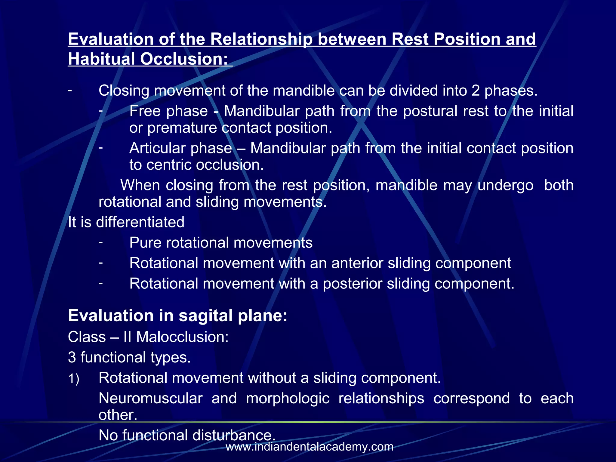
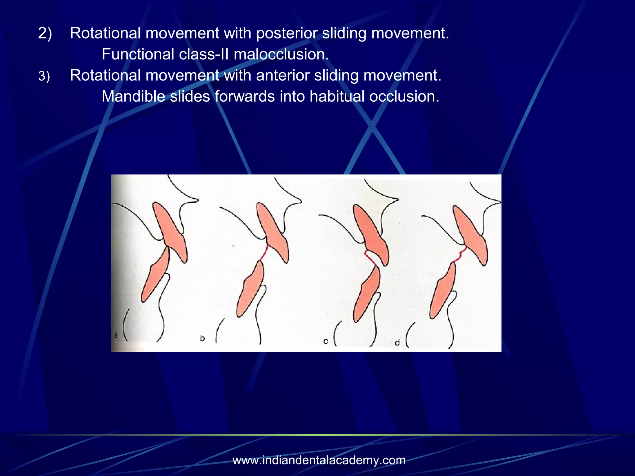
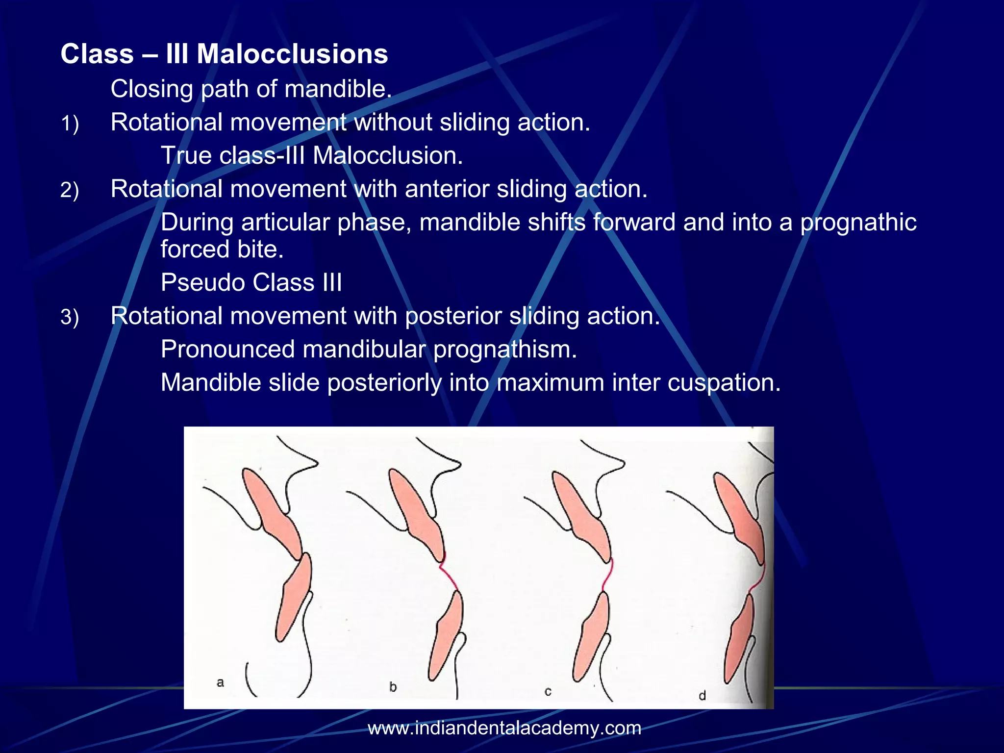
Explores postural rest position, TMJ examination, and methods for determining relationships.Details on methods to register mandibular rest position and evaluation techniques.Describes mandible movement phases and evaluations of malocclusion in different classes.
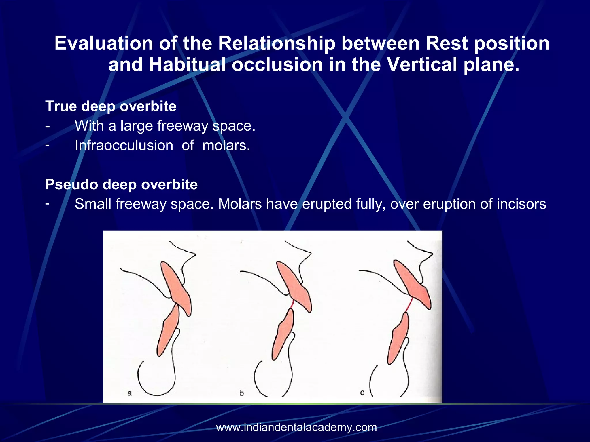
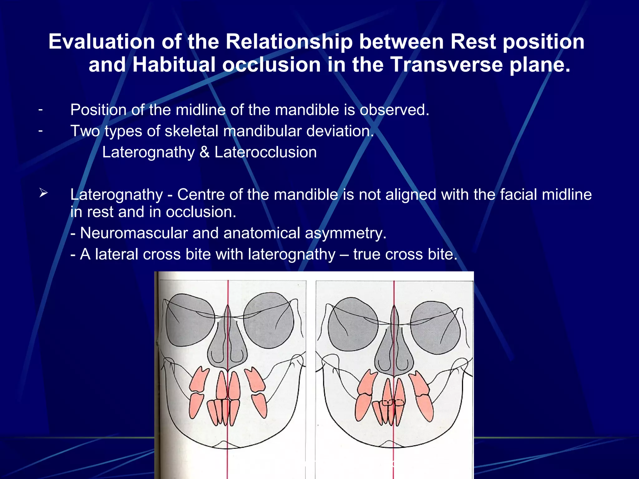
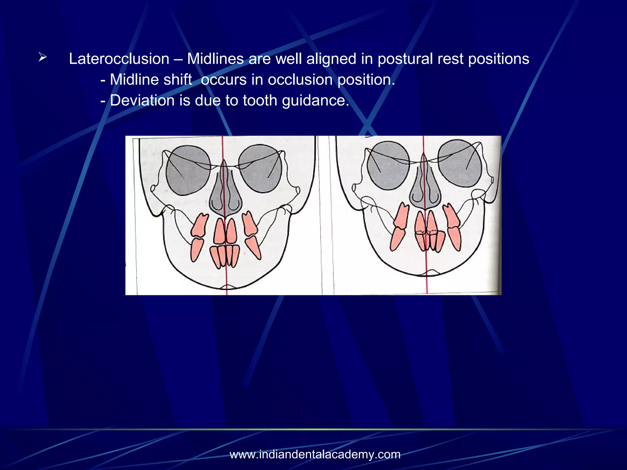
Evaluates relationships between rest position and occlusion in the vertical and transverse planes.
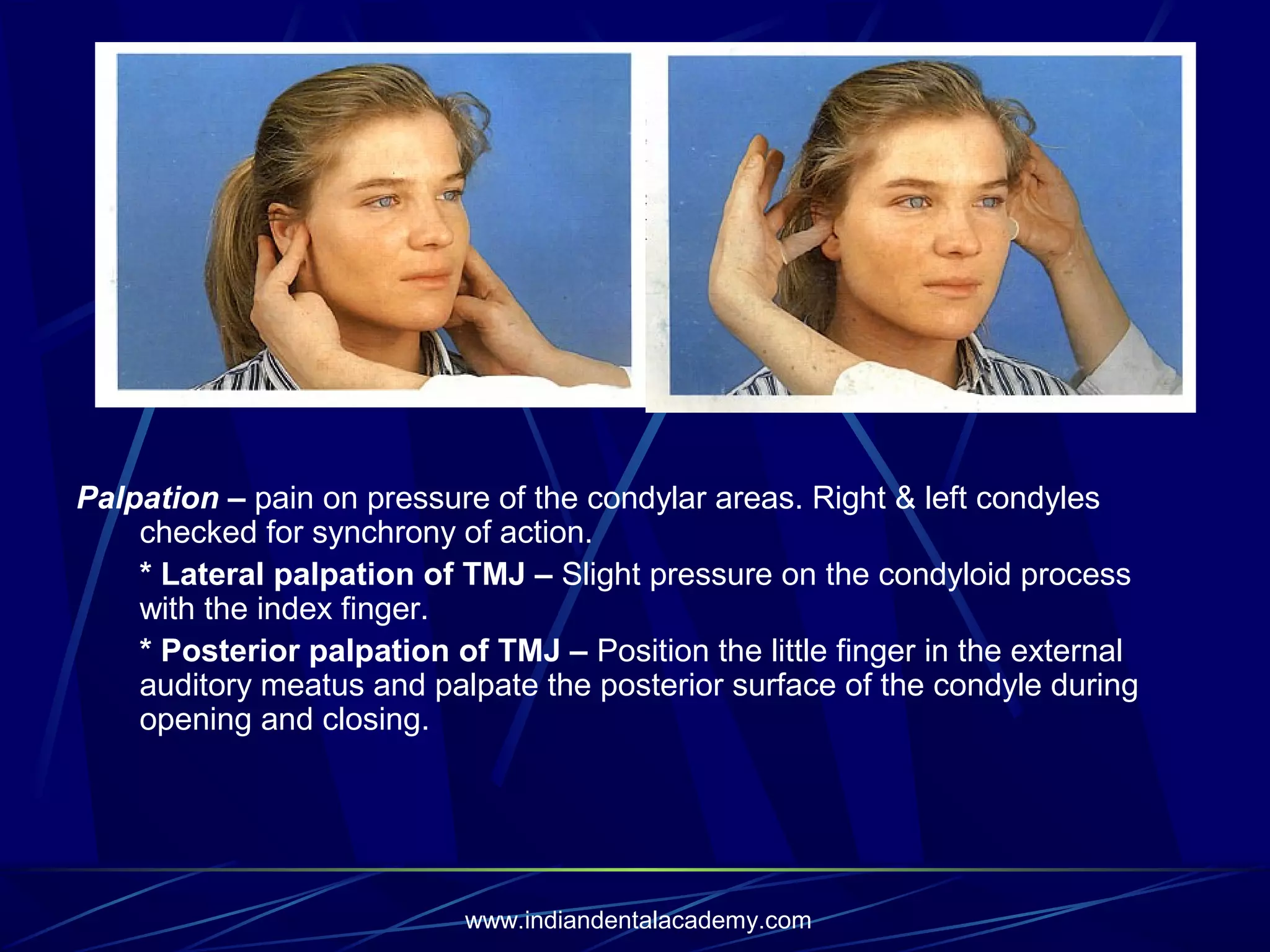
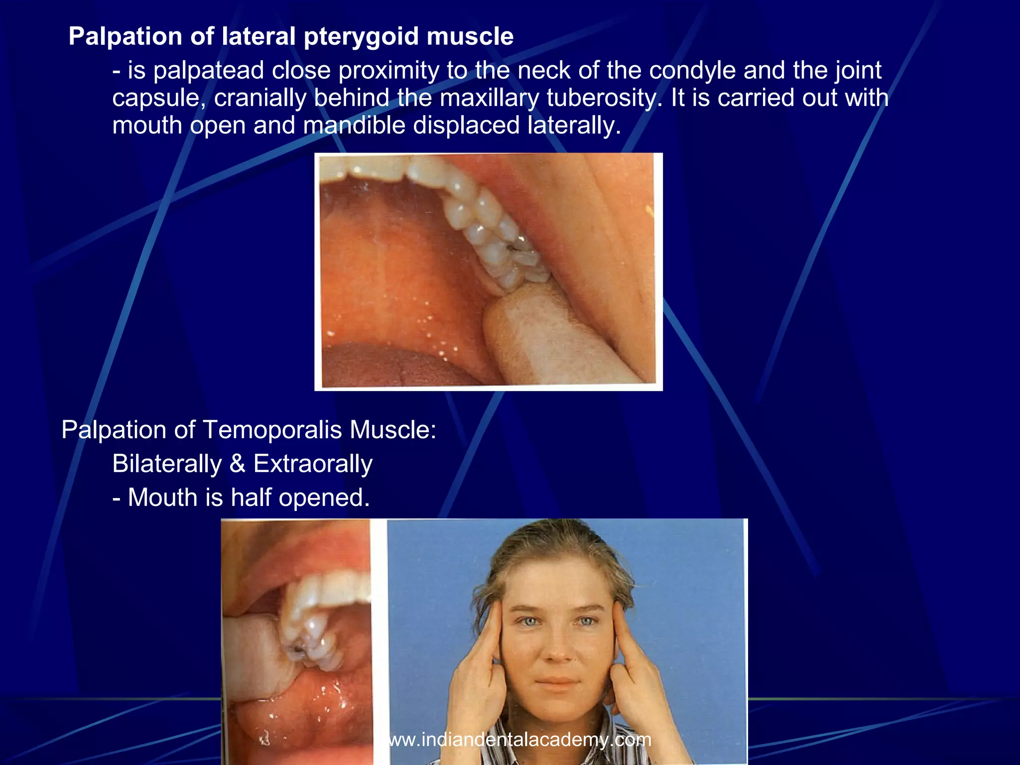
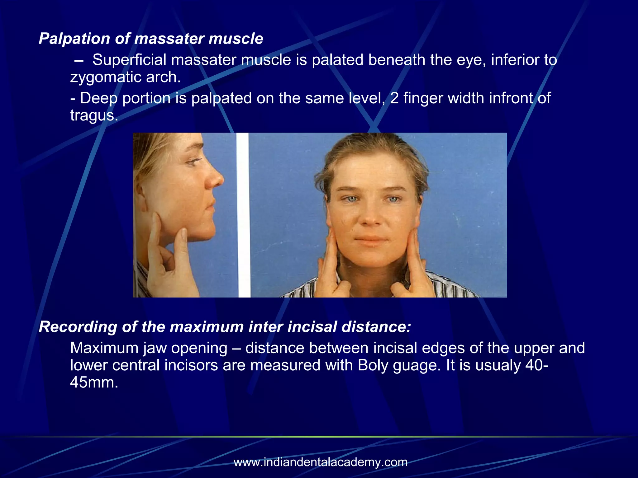
Clinical examination methods for TMJ, including auscultation and palpation strategies.
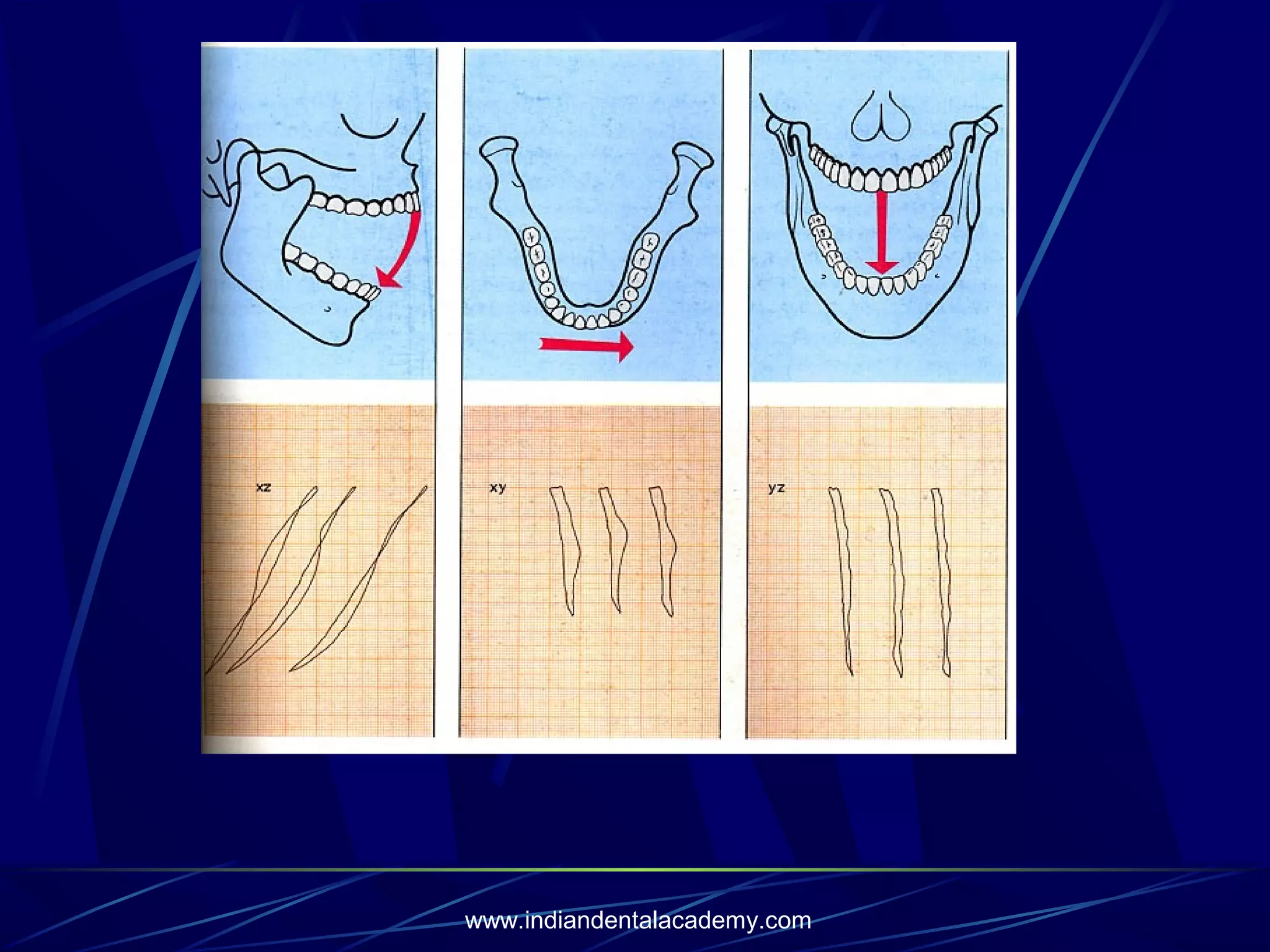
Analysis of mandibular movements and electronic registration techniques used in TMJ assessment.
Conclusion and gratitude for attending the functional analysis presentation.
Functional analysis /certified fixed orthodontic courses by Indian dental academy
1.
FUNCTIONAL ANALYSIS
INDIAN DENTALACADEMY
Leader in continuing dental education
www.indiandentalacademy.com
2.
FUNCTIONAL ANALYSIS
Examination ofthe postural rest position and
maximum intercuspation.
Examination of the temporomandibular joint.
Examination of orofacial dysfunctions.
Examination of Relationship:
Postural rest Position – Habitual Occlusion
Determination of the postural rest position.
Registration of the postural rest position.
Evaluation of the relationship postural rest position –
habitual occlusion, in three planes of space.
www.indiandentalacademy.com
3.
Determination of PosturalRest Position
Def: Relationship of the mandible to maxilla when the properly aligned
condyle disc assemblies are in the most superior position against the
eminentia, irrespective of the tooth position or vertical dimension.
-
Should be determined when the patient relaxed and sitting upright.
Frankfort horizontal plane parallel to the floor.
Tapping test can be done to relax the musculature.
When the mandible is in the postural resting position, it is usually 23mm below and behind centric occlusion.
www.indiandentalacademy.com
Registration of RestPosition:
Techniques:
Intra-oral indirect Method
(Registration with impression material)
Extra-oral direct Methods
(Registration by means of skin reference points)
- Extra-oral indirect Method
a) Roentgenocephalometric registration.
b) Kinesiographic registration
Mandibular rest position is registered 3-dimensionally.
Position of the mandible is recorded electronically.
A permanent magnet fixed with rapid setting acrylic to the lower
anterior teeth.
A Sensor system of 6 magnetometers mounted on spectacle
frames.
Every movement of the mandible and the attached magnet out of
centric occlusion, alters the strength of magnetic field. Changes
are recorded by sensors processed in the Kinesiograph and
displayed on a storage oscilloscope.
www.indiandentalacademy.com
6.
Evaluation of theRelationship between Rest Position and
Habitual Occlusion:
Closing movement of the mandible can be divided into 2 phases.
Free phase - Mandibular path from the postural rest to the initial
or premature contact position.
Articular phase – Mandibular path from the initial contact position
to centric occlusion.
When closing from the rest position, mandible may undergo both
rotational and sliding movements.
It is differentiated
Pure rotational movements
Rotational movement with an anterior sliding component
Rotational movement with a posterior sliding component.
-
Evaluation in sagital plane:
Class – II Malocclusion:
3 functional types.
1) Rotational movement without a sliding component.
Neuromuscular and morphologic relationships correspond to each
other.
No functional disturbance.
www.indiandentalacademy.com
7.
2)
3)
Rotational movement withposterior sliding movement.
Functional class-II malocclusion.
Rotational movement with anterior sliding movement.
Mandible slides forwards into habitual occlusion.
www.indiandentalacademy.com
8.
Class – IIIMalocclusions
1)
2)
3)
Closing path of mandible.
Rotational movement without sliding action.
True class-III Malocclusion.
Rotational movement with anterior sliding action.
During articular phase, mandible shifts forward and into a prognathic
forced bite.
Pseudo Class III
Rotational movement with posterior sliding action.
Pronounced mandibular prognathism.
Mandible slide posteriorly into maximum inter cuspation.
www.indiandentalacademy.com
9.
Evaluation of theRelationship between Rest position
and Habitual occlusion in the Vertical plane.
True deep overbite
With a large freeway space.
Infraocculusion of molars.
Pseudo deep overbite
Small freeway space. Molars have erupted fully, over eruption of incisors
www.indiandentalacademy.com
10.
Evaluation of theRelationship between Rest position
and Habitual occlusion in the Transverse plane.
-
Position of the midline of the mandible is observed.
Two types of skeletal mandibular deviation.
Laterognathy & Laterocclusion
Laterognathy - Centre of the mandible is not aligned with the facial midline
in rest and in occlusion.
- Neuromascular and anatomical asymmetry.
- A lateral cross bite with laterognathy – true cross bite.
www.indiandentalacademy.com
11.
Laterocclusion – Midlinesare well aligned in postural rest positions
- Midline shift occurs in occlusion position.
- Deviation is due to tooth guidance.
www.indiandentalacademy.com
12.
Examination of TemporomandibularJoint
Clinical Examination:
Auscultation – clicking and crepitus
Joint clicking is differentiated
* Initial clicking – retruded condyle in relation to disc.
* Intermediate clicking – unevenness of the condylar surfaces and of the
articular disc which slide over one another during the movement.
* Terminal clicking – condyle move to far anteriroly in relation to disc on
maximum jaw opening
* Reciprocal clicking – during opening and closing and express an
incordination between displacement of condyle and disc.
www.indiandentalacademy.com
13.
Palpation – painon pressure of the condylar areas. Right & left condyles
checked for synchrony of action.
* Lateral palpation of TMJ – Slight pressure on the condyloid process
with the index finger.
* Posterior palpation of TMJ – Position the little finger in the external
auditory meatus and palpate the posterior surface of the condyle during
opening and closing.
www.indiandentalacademy.com
14.
Palpation of lateralpterygoid muscle
- is palpatead close proximity to the neck of the condyle and the joint
capsule, cranially behind the maxillary tuberosity. It is carried out with
mouth open and mandible displaced laterally.
Palpation of Temoporalis Muscle:
Bilaterally & Extraorally
- Mouth is half opened.
www.indiandentalacademy.com
15.
Palpation of massatermuscle
– Superficial massater muscle is palated beneath the eye, inferior to
zygomatic arch.
- Deep portion is palpated on the same level, 2 finger width infront of
tragus.
Recording of the maximum inter incisal distance:
Maximum jaw opening – distance between incisal edges of the upper and
lower central incisors are measured with Boly guage. It is usualy 4045mm.
www.indiandentalacademy.com
16.
Opening and closingmovements of mandible
‘C’ and ‘S’ type of deviation are typical signs of functional disturbance.
Registration of the mandibular movement
- Electronic recording unit
A magnet is placed intraorraly on the mandible to produce a three
dimensional magnetic field. Head frame consists of a system of antennas
which record the changes in position of the magnetic field during
movement of the mandible. The signals are converted to analog electrical
charges which are fed into a processor and displayed as three reference
values. X, Y & Z.
Graphically represented in XY plane – horizontal plane
YZ frontal plane
XZ – sagital plane
TMJ – Radiographic Examination
- Position of the condyle in relation to fossa
- Width of joint space.
- Changes in shape and structure of condylar head
- Mandibular fossa.
www.indiandentalacademy.com